Diabetes Pakistan Metabolic Syndrome Date: 1st - 2nd November, 2024 Venue: Serena Hotel, Faisalabad
For Emergencies: +92 41-2694037

Red Light Therapy Benefits: A Complete Guide to Health, Skin, and Wellness

Dr. Ahmad Shahzad
Founder | Lyallpur Diabetes Foundation
Consultant Diabetologist | Educator | Advocate for Preventive Care

Red light therapy is among the most discussed therapies in the world of health and wellness. This non-invasive approach has numerous possible benefits, starting with making the skin look better and ending with relieving muscle pains. With support of the increasing research and real life stories of success, knowing red light therapy benefits will prove helpful in deciding whether it is the right self-care or recovery option of your routine.

What is Red Light Therapy?

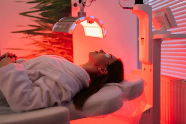
Red light therapy (RLT) is a non-invasive therapy involving low-wavelength red and near-infrared light to benefit the skin, wounds, and inflammation. The treatment is effective because it activates the mitochondria, the powerhouses of body cells, to produce greater energy. 

NASA initially developed the technology to cultivate crops in space and cure astronauts.

How red light therapy works

In an RLT session, you have your skin subjected to a machine that gives out low-energy red light. The light penetrates the skin to the superficial level (1 to 10 millimeters). As the cells absorb this energy in the light, it activates the mitochondria to generate more adenosine triphosphate (ATP), the energy carrier in cells. This cellular energy is believed to stimulate cell function, repair, and regeneration. 

Common uses and potential benefits

Although there are promising studies, other experts indicate that additional research is required in order to establish the complete efficacy of RLT. 

Widely used red light therapy is:

  • Skin rejuvenation: RLT is commonly advertised as a way to treat the skin. It can trigger the production of collagen and elastin, which helps minimize fine lines and wrinkles. It may be used even in the case of sun damage, uneven color, and acne.
  • Wound healing: By promoting blood circulation and stimulating cell repair, RLT may enhance wound healing, burn healing, and scar healing.
  • Hair growth: RT has been found to induce hair follicles in individuals with androgenic alopecia (hereditary hair loss), leading to new growth and a higher level of thickness.
  • Pain and inflammation: Other studies indicate that RLT may be able to treat pain and inflammation related to various conditions, such as arthritis, muscle soreness, and joint pains.
  • Neurological health: Neurological studies are looking at using red light to enhance the cognitive ability of individuals with dementia because the light has the potential to increase blood flow and oxygen to the brain.
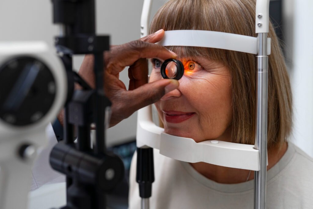
Safety and devices

Non-UV light is used in RLT, hence, it does not burn the skin or elevate the risk of skin cancer linked to UV radiations. It is also thought to be safe when used in short term and it is not invasive. The side effects are mild and short term like skin irritation. 

The administration of RLT may use a number of devices:

  • Professional equipment: More powerful equipment is employed in medical practice by dermatologists and other medical caregivers.
  • Home units: Masks, wands, and panels can be used at home, but are frequently less effective than professional. To be safe, make sure the device is FDA-cleared and always put on protective eyewears as directed.

How to Use Red Light Therapy Effectively

Red light therapy requires you to focus on targeted, regular treatments with the correct device to achieve the desired results. A typical routine of 3-5 sessions a week, each 10-20 minutes, is important in achieving results in a few weeks or months. 

For at-home use

If you are using a consumer-grade red light therapy device, follow these best practices for effective treatment: 

  • Choose the right device. Select a device based on your treatment goals.
    • For skin issues, like wrinkles or acne on your face, a mask or handheld wand is best.
    • For pain or hair loss, a portable device, flexible wrap, or panel can target specific areas of your body, like a knee or the scalp.
  • Choose the important specifications. Find equipment that employs a particular therapeutic wavelength, 660 nanometers (red light) and 850 nanometers (near-infrared). Greater power output (irradiance) may result in more successful shorter sessions.
  • Prepare the area. Wash your skin and take off all makeup, dirt or oil before treatment. In skin problems and loss of hair, the light should be used on bare skin.
  • Position the device correctly.
    • For surface-level skin treatment, devices should generally be positioned 12 to 36 inches away.
    • For deeper issues like muscle and joint pain, move the device closer, typically 6 to 12 inches from the skin.
  • Keep up to a regular routine. Apply 10- 20 minutes, 3-5 times per week. The time-consistency is preferable to longer, individual sessions. It can take 2 to 4 weeks before results of some conditions can be seen.
  • Protect your eyes. Wear the eye protection that is provided with your device, particularly when you are aiming at your face, or when you are facing the light itself. 

Considerations for specific goals

Your goals may require specific routines or a combination of therapies. 

  • To rejuvenate the skin: Be patient, and it can take weeks of regular use before one experiences the benefits of reduced wrinkles or better tone. Addressing this, it is possible to enhance the benefits by combining it with a good skincare regimen.
  • To relieve pain and recover muscles: Red light therapy is used by many athletes prior to and after exercise to prevent injuries and accelerate healing. In chronic pain, a higher frequency of sessions (45 times per week) can be useful during flare-ups.
  • To grow hair: It is crucial to be consistent and it can be up to 3 to 6 months before you see an improvement. Red light therapy can be used with other hair growth therapies such as minoxidil or microneedling.

Maximize results with supportive habits

To support the effectiveness of red light therapy, consider incorporating these wellness practices: 

  • Maintain a healthy diet and stay hydrated.
  • Manage stress and ensure good sleep hygiene.
  • Include regular exercise in your routine. 

When to consult a professional

Home appliances are generally not as powerful as professional devices and can be successful. Never begin a new therapy, particularly one that is topical, without consulting a healthcare provider or dermatologist, particularly when you have sensitive skin, photosensitivity or underlying medical issues.

You may also like to read: What is Tdap Vaccine?

Final Thoughts

The list of benefits of red light therapy is quite broad, including the improvement of the skin condition and hair growth, pain reduction, and overall wellness. Although not a miracle, long-time and safe usage can provide significant improvements to many individuals. When you know the established positive effects of red light therapy and apply the correct method, it will become a worthwhile addition to your health and self-care regimen.

FAQs

Who should not use red light therapy?

Consult your doctor first in case you have lupus, a light-sensitive condition, or are on drugs that predispose you to sunlight sensitivity (e.g. antibiotics or retinoids). Only devices that are flicker free and endorsed by the neurophysician will work with epileptics.

Is 10 minutes of red light therapy enough?

Frequent red light therapy sessions, 3 to 5 times per week can aid skin, muscle and pain relief objectives. Once visible results are achieved maintenance sessions (one to two times a week) are used to maintain the long-term benefits. Ten to twenty minutes of sessions long are effective, and can be easily sustained.

How do I know if red light therapy is working?

Red light therapy is fast acting on acute problems, like an injury, and is slow acting on chronic problems, like arthritis. The condition of many patients improves directly after the initial red light therapy session and improves during the next several weeks.

What is Tdap Vaccine? Benefits, Schedule, and Everything You Need to Know

Dr. Ahmad Shahzad
Founder | Lyallpur Diabetes Foundation
Consultant Diabetologist | Educator | Advocate for Preventive Care

Vaccines play a vital role in protecting us from serious illnesses, and one of the most important among them is the Tdap vaccine. If you’ve ever wondered what is Tdap vaccine and why it matters, the answer lies in its ability to guard against three dangerous diseases—tetanus, diphtheria, and pertussis. The vaccine is recommended to adolescents, adults, and even in pregnancy, not only does it protect individuals but communities as well.

What is Tdap Vaccine?

Tdap vaccine is an immunization that combines against tetanus, diphtheria, and pertussis (whooping cough). It is targeted at adults, teens, and children of an older age. The vaccine resembles the DTaP vaccine used in infants and younger children, except that it administers less dose of the diphtheria and pertussis antigens. 

What Tdap protects against

  • Tetanus: Tetanus is a serious infection typically caused by bacteria commonly found in soil, dust, and manure. It comes into the body through cuts or open wounds, and it causes a poison that leads to painful muscle contractions.
  • Diphtheria: This illness primarily affects the throat and nose, producing a toxin that can lead to severe health complications, including heart failure and breathing difficulties. The bacteria are contagious.
  • Pertussis (Whooping Cough): A very infectious respiratory disease that may be especially risky in infants and young children. It can result in severe coughing fits, vomiting, and cracked ribs, although it may result in milder illness in older people. 

Who should get the Tdap vaccine

  • Adolescents: Tdap should be used as a single dose in adolescents aged 11 or 12 years.
  • Adults: All adults 19 and above who have not had a Tdap booster are to receive one. One Tdap or Td booster is advised every 10 years after the first dose.
  • Pregnant individuals: The vaccine is recommended at every pregnancy, preferably at a gestation of between 27 and 36 weeks. This is to transfer protective antibodies to the baby to protect against whooping cough during their first few months of life, when they are at the highest risk.
  • Individuals with wounds: It can be administered to manage the wounds, provided that at least five years since the previous tetanus vaccine. 

 Tdap Vaccine Schedule and Boosters

 Tdap Vaccine Schedule and Boosters

The Tdap vaccination schedule has one dose in adolescents, a booster each after 10 years in adults, and one dose in each pregnancy. Severe wounds might also necessitate a Tdap shot or to have a back up on missed vaccinations. 

Tdap for adolescents

Tdap should be given once in adolescents, normally in the age group of 11 or 12 years. This Tdap dose is used instead of one of the DTaP vaccines of the childhood immunization series. 

Tdap for adults

Booster shots should be taken every 10 years in adults. Following the first dose of Tdap, you should get either a Tdap or Td (tetanus and diphtheria) booster. The Tdap booster is best as it offers protection against whooping cough. 

Tdap during pregnancy

  • The Centers of Disease Control and Prevention (CDC): suggests a single dose of Tdap in all pregnancies in pregnant individuals. 
  • Timing: The ideal time is to administer the shot in the third trimester, ideally in 27 to 36 weeks of gestation.
  • Purpose: The vaccine helps the mother’s body to generate the protective antibodies, which are transferred to the baby before birth. It will offer the much-needed temporary immunity to whooping cough in the first weeks of the infant’s life when they are the most susceptible.
  • Vaccination in all pregnancies: With time, the amount of antibodies lost, so it is necessary to do the vaccination each time, giving maximum protection to the child born. 

Tdap for wound management

In case of a severe cut or wound, this may require the tetanus-containing vaccine based on your immunization history. 

  • In case you have never received a Tdap shot previously, you will be given one to treat wounds.
  • When you already receive Tdap, a Tdap or Td booster is advised when it is over five years since the last injection.

Catch-up schedule

For individuals aged 7 and older who were never vaccinated or did not complete the DTaP series as children, a three-dose catch-up series is recommended. 

  • At least one of these doses should be Tdap.
  • The remaining doses can be either Td or Tdap. 

Benefits 

This is especially crucial for safeguarding infants and other vulnerable populations from serious illness. 

  • Avoids life-threatening tetanus (lockjaw): Tetanus is a severe bacterial infection that may enter the body through a cut or a wound and causes painful muscle spasms that may cause breathing problems, paralysis, and death. The Tdap vaccine triggers the immune system to generate antibodies against the tetanus toxin, which prevents the disease.
  • Prevents complications of diphtheria: Diphtheria is another contagious disease that forms a thick layer in the throat that may block breathing. The diphtheria component of the vaccine is against severe respiratory symptoms and complications such as heart failure, nerve damage, paralysis, and death.
  • Lessens the severity of whooping cough: The protection by the pertussis vaccine may decrease with time, but it is very efficient in stopping severe symptoms and complications. In adults who become infected with whooping cough following vaccines, the disease is milder and less lasting.
  • Prevents whooping cough during early infancy: Women who take Tdap vaccine during pregnancy transfer protective antibodies to their unborn babies. This is the only means of protecting the babies against whooping cough in the first few months of their lives when they are too young to be inoculated and most susceptible to serious, life-threatening complications.

Common side effects

Most individuals receiving the Tdap vaccine show no side effects or minor ones that clear off within a few days. The side effects most frequently occurring are: 

  • Pain, redness, or swelling where the shot was given
  • Mild fever
  • Headache
  • Fatigue
  • Nausea, vomiting, diarrhea, or stomachache 

You may also like to read: Stem Cell Therapy for Diabetes

Tdap vs. DTaP: What’s the Difference? 

The difference between the Tdap and DTaP vaccines lies in their targeted age group and the dosage of the diphtheria and pertussis components. Both are combination vaccines that protect against tetanus, diphtheria, and pertussis (whooping cough). 

Feature DTaP VaccineTdap Vaccine
RecipientsInfants and young children under 7 years old.Adolescents (11–12 years) and adults.
PurposeAdministered in a series of shots to build strong, foundational immunity in a child’s developing immune system.Used primarily as a booster to maintain protection, as childhood immunity wanes over time.
DosageContains a full-strength dose of diphtheria and pertussis components.Contains a full-strength tetanus dose but a reduced dose of diphtheria and pertussis components. The lowercase “d” and “p” in “Tdap” denote these smaller doses.
SchedulePart of the routine childhood immunization series, typically given in five doses at 2, 4, and 6 months, 15–18 months, and 4–6 years.A single dose is given around 11–12 years of age, followed by a booster every 10 years throughout adulthood. It is also given during every pregnancy.

The “acellular” component

The two vaccine names contain the a which is used to denote acellular. This implies that the vaccine would not require the entire pertussis bacterium, but a few components of it to produce an immune response. This is done to minimize the side effects of previous pertussis vaccines that were made from whole cells.

Final Thoughts

Knowledge of what is Tdap vaccine is emphasizes its significance in the prevention of tetanus, diphtheria, and pertussis. By keeping abreast with Tdap vaccinations, people are safeguarding themselves, as well as protecting those most at risk, namely infants and the elderly. Having this vaccine in your health plan is an easy but effective way of protecting yourself long-term and healthy society.

FAQs

How often is a Tdap shot needed?

Adults. CDC advises vaccination against diphtheria every 10 years in all adults. After an adult has a dose of Tdap, use Td or Tdap as a booster dose.

Is a tetanus shot the same as Tdap?

Td vaccine only protects against tetanus and diphtheria. Tdap is used to prevent tetanus, diphtheria and pertussis (whooping cough). Whooping cough is particularly harmful to young children and infants below the age of 6 months.

Who should not get the Tdap vaccine?

Has received an allergic reaction following a prior dose of any vaccine against tetanus, diphtheria, or pertussis, or has any life threatening severe allergies.

The Importance of Healthy Eating for Metabolic Health

Dr. Ahmad Shahzad
Founder | Lyallpur Diabetes Foundation
Consultant Diabetologist | Educator | Advocate for Preventive Care

Having a healthy metabolism is a key to general wellbeing. It determines the way the body utilizes energy, glucose levels, and essential body functions. Healthy eating has been one of the best ways to safeguard and enhance metabolism health. The type of foods we eat daily is strong enough to balance hormones, add energy and decrease the chances of developing diseases. Through a healthy, balanced diet, we can boost our metabolism and establish an improved health base in the future.

Understanding Metabolic Health

Metabolic health is defined as a normal operation of your body metabolism, the way of how it uses the food to produce energy. Not only does it concern weight, but a whole-body examination of how effectively your body utilizes and stores energy. Being in good metabolic health may reduce your chances of having chronic diseases like type 2 diabetes, heart disease and stroke.

Key indicators of metabolic health

Medical workers consider a variety of biomarkers. They are usually measured during a general physical check-up to determine your metabolic health:

  • Blood sugar: The normal level of blood sugar in the body means that you have enough insulin to break down sugar into energy. Fluctuating levels may be an indication of insulin resistance.
  • Waist circumference: Fat around the belly (an apple shape) is a major risk factor of metabolic syndrome. A set of diseases that puts you at risk of heart disease and diabetes.
  • Blood pressure: Having high blood pressure on a chronic basis can hurt your heart and blood vessels.
  • Triglycerides: A large amount of these blood fats may put you at risk of heart disease.
  • HDL (good) cholesterol: Low amounts of HDL cholesterol levels can predispose you to heart disease.

A combination of three or more of these risk factors can indicate metabolic syndrome. 

How Nutrition Affects Metabolism

Nutrition directly affects your metabolism by influencing your energy intake and expenditure, regulating blood sugar, and providing the necessary building blocks for all of your body’s processes. The quality and type of food you eat can either optimize or impair your metabolic health. 

The role of macronutrients

  • Carbohydrates: Your body’s primary energy source.
    • Metabolic effect: The nature of the carbohydrate is important. Uncomplicated, complex carbohydrates (such as sugary beverages, white bread) induce fast glycemic surges, which could result in insulin resistance and type 2 diabetes. Complicated carbohydrates in whole grains, fruits, and vegetables maintain a slower and more steady discharge of energy.
  • Proteins: Proteins, which are vital in making and repairing tissues, possess a greater “thermic effect,” or your body expends more energy to digest it than fats and carbs.
    • Metabolic effect: Lean protein diet can temporarily increase your metabolism and retain muscle mass to avoid the metabolic slowdown that is accompanied by weight loss.
  • Fats: While fat has the highest energy density, the type of fat consumed is critical.
    • Metabolic effect: Unsaturated fats (nuts, seeds, oils, etc.) may enhance the metabolic health, decrease inflammation and increase insulin sensitivity. In contrast, too much saturated and trans fats may lead to metabolic disorders. 

The impact of food processing

  • Highly processed foods, such as fast food, packaged snacks, and sugary drinks, are detrimental to metabolic health.
    • Nutritional deficiencies: Processing usually takes away healthy nutrients such as fiber, vitamins, and minerals and imparts too much sugar, salt, and unhealthy fats.
    • Fast digestion: Processed food is very rich in refined carbohydrates and low in fiber, thus digestible quickly, which results in blood sugar surges and crashes.
    • Damage to gut health: Processed foods can ruin your gut microbiome, and unbalanced microbiome is associated with metabolic dysregulation. 

The importance of micronutrients and fiber

  • Micronutrients (vitamins and minerals) are vital for metabolic function, acting as cofactors for enzymes involved in energy production.
    • Examples: Deficiency of vitamin D is associated with metabolic syndrome and such minerals as magnesium and zinc are important in insulin sensibility.
  • Dietary fiber plays a key role in metabolic health, primarily by feeding beneficial gut bacteria.
    • Metabolic effect: Fiber fermentation by intestinal microbes gives rise to short-chain fatty acids that play a wide range of physiological functions that can enhance insulin sensitivity, decrease inflammation, and aid in the regulation of appetite. 

Healthy Eating Habits for Better Metabolic Health

Healthy Eating Habits for Better Metabolic Health

Healthy eating habits form a foundation of enhancing metabolic health through efficient control of blood sugar, weight management, and prevention of metabolic-related chronic illnesses. What, when, and how you consume food will enable you to maximize the utilization of energy and metabolism in your body.

Focus on nutrient-dense, whole foods

  • Make protein a priority: Add lean protein foods such as fish, eggs, beans and chicken to all the meals. Protein aids in regulating blood sugar, it enhances satiety, and it preserves muscle mass which is a significant glucose consumer.
  • Load up on fiber: Foods rich in fiber, including vegetables, fruits, and whole grains, aid in controlling blood sugar and maintaining a healthy microbiome of the gut. Fiber can make you fuller as well which can combat overeating.
  • Practice healthy fats: Include healthy fats such as avocados, nuts, seeds and olive oil. Healthy fats enhance sensitivity of Insulin and may lower insulation which helps in metabolic health. 

Be mindful of meal timing

·       Eat more often: Time you’re eating behaviors according to the circadian rhythm of your body and eat most of your calories during the morning. High-calorie breakfast is associated with more weight loss and better glucose and insulin responses than high-calorie dinner.

·       Eat regular-balanced meals: Frequent, balanced meals keep blood glucose levels constant and eliminate energy crashes and overeating. Missing meals would slow down your metabolic rate.

·       Limit nighttime eating: Do not eat late at night, late night eating has been associated with glucose intolerance, fatteries and weight gain.

Practice mindful eating and portion control

  • Eat consciously: Be aware of the eating experience, enjoy the taste and the texture. It could assist you in becoming aware of the natural signals of hunger and fullness in your body and avoid overeating.
  • Control portions: Half of your plate of vegetables can help you control the size of portions. Conscious eating enables one to control the sugar and hormone levels in the blood that determine the feeling of fullness.
  • Meal prep: Plan and pre-cook meals ahead of time to stay in charge of nutrition and not resort to unhealthy and impulsive food when you are in a hurry. Meal prepping is also beneficial in terms of portion control and consistency.

You may also like to read: Prenatal Vitamins

Consequences of Poor Eating on Metabolism

Mistresses diet is damaging to your metabolism and usually results in a variety of chronic health problems. Diet rich in processed foods and sugar, low in fiber and consumed at unusual hours may essentially disturb the metabolism in the body.

The consequences of a poor diet

  • Weight gain: Overconsumption of calories particularly sugary beverages and processed foods can cause weight gain and become obese. This overweight is a major risk factor towards metabolic syndrome especially around the abdomen.
  • Frequent spikes in blood sugar: High-refined carbohydrate diets and the addition of sugars lead to insulin resistance and type 2 diabetes. In the course of time, the cells of your body might become less sensitive to insulin, and it is known as insulin resistance. This may cause type 2 diabetes, when not controlled.
  • Liver is damaged and fats: The liver processes fructose, a kind of sugar that is common in processed food and sugary drinks the way it processes alcohol. The abundance of fructose may increase the fat level in the liver which is part of non-alcoholic fatty liver disease, a predisposing factor to diabetes and heart disease.
  • Gut microbiome dysbiosis: The unequal diet, especially high in sugar and unhealthy fats, can disrupt the balance of your gut bacteria. This dysbiosis may decrease useful bacteria and enhance inflammatory types, which lead to metabolic imbalances and chronic and low-grade inflammation.
  • Avoiding regular eating patterns: Missing meals or having late meals are known to disrupt the metabolism of your body. When you spend extended time without meals the body must be in survival mode and as a result, your metabolism will decrease, and the energy will be stored. Abnormal eating has also been attributed to increased insulin resistance and metabolic syndrome.
  • Low activity and tiredness: A fluctuating level of blood sugar due to a large consumption of simple sugars may cause the phenomenon of a high and low energy level. This impacts your vitality, mood and state of mind. 

Impact on the metabolic cycle

The metabolic cycle is influenced by food quality and consistency. 

  • Diet with a high level of fiber and nutrients with regular eating time helps in maintaining a balanced metabolism.
  • Poor intake of fiber and excess sugar, infrequent eating leads to disrupted metabolic signaling, which causes weight gain, resistance to insulin and systemic inflammation.

Bottom Line

To sum up, proper nutrition is a key to good metabolic activity, as it allows the body to work effectively and reduces the chances of chronic illnesses. Through wholesome, nutrient-rich foods and mindful eating, we will be able to contribute to energy balance, enhance long-term well-being, and resilient metabolism. Even minor changes in the daily meal can have long term effects of benefit to health and vitality.

FAQs

Q: How do I understand metabolic health?

Metabolic health is characterized by the optimal level of 5 clinical markers blood sugar, triglycerides, high-density lipoprotein (HDL) cholesterol, blood pressure, and waist circumference. The individual values as well as the variability of these markers are important in helping to decode your metabolic health.

Q: What is the biggest metabolism killer?

Overall, high saturated fats, sodium, and added sugars, coupled with low antioxidants, fiber, vitamins, and minerals are the worst foods to have regarding your metabolism. These foods (and beverages) may elevate the exposure to inflammatory and oxidative stress, which preconditions ill metabolic health.

Q: How to reset metabolism after 40?

  1. Eat at regular times.
  2. Eat enough calories.
  3. Eat more protein.
  4. Drink green tea.
  5. Lift weights.
  6. HIIT.
  7. Drink more water.
  8. Manage stress.

Q: Can sleep affect your metabolism? There are increased research and interest that sleep disorders and sleep loss affect metabolism. Lab research has made it very clear that sleep deprivation has the capability of modifying the glucose metabolism and hormones in metabolism regulation, that is, the reduction in leptin and the increase in ghrelin.

Treating Diabetic Retinopathy: Options and Outcomes

Dr. Ahmad Shahzad
Founder | Lyallpur Diabetes Foundation
Consultant Diabetologist | Educator | Advocate for Preventive Care

One of the eye complications that are commonly associated with diabetes and a major cause of vision loss throughout the world is diabetic retinopathy. Unattended, it may silently develop and destroy the sensitive blood vessels in the retina causing blurred vision or even blindness. Luckily, there are some useful means to treat and cope with this disorder using modern medicine. We shall briefly examine the various treatment opportunities that exist with diabetic retinopathy in this article and how patients can hope to achieve once care is obtained in time.

What is Diabetic Retinopathy?

Diabetic retinopathy is the most frequent diabetic complication that impairs blood vessels of the retina, the light sensitive tissue at the back of the eye, which is the main cause of vision loss and blindness.

Treatment Options for Diabetic Retinopathy

Diabetic retinopathy treatment depends on stage and severity of the disease but includes management of blood sugar to highly complex surgical interventions. It is aimed to stop the development of the disease and avoiding further loss of vision because in many cases, previously damaged parts of the eye are irreversible.

Medical management

In the case of early-stage diabetic retinopathy, an ophthalmologist can only prescribe routine follow-up and rigid control of your general condition.

  • Diabetes management: This is through strict regulation of your blood sugar level, which will prevent or at least delay the disease.
  • Blood pressure and cholesterol: High blood pressure and high cholesterol can be treated and help to lessen the risk of the worsening of retinopathy.
  • Fenofibrate: In individuals with non-proliferative diabetic retinopathy (NPDR), a cholesterol-lowering drug known as fenofibrate has been proven to decrease the chances of the condition progressing even those with normal lipid levels. 
Treatment Options for Diabetic Retinopathy

Eye injections

The swelling of the macula (central part of the retina) and growth of new, fragile blood vessels is treated with injections of medication directly into the eye to prevent the disease.

  • Anti-VEGF drugs: These encompassed Avastin, Eylea, Lucentis, and Vabysmo, which are anti-vegetable endothelial growth factor (VEGF) drugs, which enlarges and permeabilizes blood vessels. One of their uses is as a first-line therapy of diabetic macular edema (DME) and proliferative diabetic retinopathy (PDR).
  • Corticosteroid implants – In cases where there is no response to anti-VEGF therapy, steroid drug could be injected or implanted into the eye and the drug would be slowly released over a few months.
  • Refillable eye implants: Newly approved models, including Susvimo, are provided with a refillable implant that pumps a specific level of a customized formulation of ranibizumab to the eye. 

Laser treatments

Laser treatments can seal leaking blood vessels and shrink abnormal new ones. 

  • Focal or grid laser: This is done in the treatment of DME. It uses laser burns that are directed at ruptured blood vessels in the macula.
  • Scatter (panretinal) photocoagulation: It is applied to cases of diabetic retinopathy which is advanced and proliferative. The laser produces small, scattered burns to the peripheral retina which shrink and disappear into the abnormal new blood vessels.

Eye surgery (Vitrectomy)

Surgery is typically reserved for advanced cases where other treatments are not sufficient. 

  • When is it needed? A vitrectomy can be used in case of severe bleeding in the vitreous gel which fails to clear, the presence of a lot of scar tissue pulling the retina, or a detached retina.
  • What happens during surgery? The surgeon takes out the hazy vitreous gel and a scar tissue and puts in a clear saline solution or a gas or oil bubble to cushion the retina. 

Emerging therapies

Ongoing research is exploring new and more effective treatments for diabetic retinopathy, including: 

  • New anti-angiogenic drugs
  • Oral medications
  • Stem-cell therapy
  • Gene therapy

Although the damage cannot be reversed, treatment can help prevent or delay additional damage to the vision. Having an integrated approach and a high level of diabetes control is usually the best course of action.

You may also like to read: Preventing Vision Loss from Diabetic Retinopathy

Expected Outcomes by Treatment Type

Treatment of diabetic retinopathy aims at preventing or halting the disease progression and to avoid severe loss of vision. The level and form of your condition, the method of treatment, and your health, in general, blood sugar control, are among the factors that define the exact outcome you can expect. Although it can be improved, particularly when the disease is diagnosed in the early stages, in most cases, it is impossible to undo all the harm.

Expected outcomes by treatment type

  • Often improve vision significantly in diabetic macular edema.
  • Results are best with consistent and ongoing treatment.
  • Reduces the risk of severe vision loss by more than 50%.
  • May cause side effects like reduced night or peripheral vision.

Factors influencing long-term outcomes

The long-term prognosis is heavily influenced by several factors, many of which you can actively manage.

  • Treatment adherence: It is important to adhere to your treatment program because under-treatment is one of the leading causes of a poor outcome in the real world.
  • Management of diabetes: Glycemic control (HbA1C), blood pressure control and cholesterol control divert renal injury disease progression and enhance treatment results.
  • The severity of the disease at diagnosis: the later the diabetic retinopathy is diagnosed and treated, the better the outcome is. Delaying action after the vision has been seriously affected may cause permanent structural harm.
  • The basic retinal damage: The treatment can correct fluid and bleeding; however, the visual prognosis is constrained by the level of irreparable harm to the photoreceptors and retinal nerve cells that might have taken place.

Final Thoughts

Therapy of diabetic retinopathy is aimed at reducing the course of the disease, preserving the vision, and enhancing the quality of life. Although the results are not identical at each phase of the condition and different types of treatments are applied, early diagnosis and regular treatment can significantly help in saving vision. Medical therapy together with proper management of diabetes can help patients deliver positive long-term outcomes and minimize chances of losing their sight severely.

FAQs

Q: What is the 4 2 1 rule for diabetic retinopathy?

The diagnosis is based on the 4-2-1 rule. Diagnosis is made when the patient has, in 4 quadrants, diffuse intraretinal hemorrhages and microaneurysms, in 4 quadrants, venous beading, and in 1 quadrant, IRMA.

Q: Can mild diabetic retinopathy be reversed?

Treatment may delay or prevent the progression of diabetic retinopathy, but it is not a cure. Diabetes is a long-term health condition and as a result, future retinal injury and vision impairment can still occur. Regular eye exams will be required even after diabetic retinopathy is treated. There may be a time you require supplementary treatment.

Q: What is the new treatment for retinopathy?

Basel, 22 May 2025 – Roche (SIX: RO, ROG; OTCQX: RHHBY) has announced that Susvimo® (ranibizumab injection) 100 mg/mL is now approved by the US Food and Drug Administration (FDA) in the treatment of diabetic retinopathy (DR), a potentially blinding disorder, which is present in nearly 10 million individuals in the USA.

Preventing Vision Loss from Diabetic Retinopathy

Dr. Ahmad Shahzad
Founder | Lyallpur Diabetes Foundation
Consultant Diabetologist | Educator | Advocate for Preventive Care

One of the most prevalent complications of diabetes and the major cause of vision loss in the globe is diabetic retinopathy. It arises when excess sugar harms the tiny blood vessels in the retina and it slowly impairs the vision. The fact that sight impairment in diabetic retinopathy may be averted in most cases through management of diabetes, eye checkups, and healthy lifestyle is good news. In this article, we’ll explore effective strategies for preventing vision loss from diabetic retinopathy and protecting long-term eye health.

Understanding Diabetic Retinopathy

Diabetic retinopathy is a disease of the eye that is caused by diabetes and is caused by the destruction of blood vessels in the retina. It is a major cause of blindness in adults and may occur in all those with Type 1, Type 2 or gestational diabetes.

Why Prevention is Crucial

Disease is important in preventing diabetic retinopathy, which is asymptomatic at its onset and causes permanent loss of vision when not treated. Protect your sight by detecting diabetes early and managing its risk factors proactively.

Benefits of prevention

  • Avoids permanent vision loss: Treatments could delay the development of advanced diabetic retinopathy, but not permanent damage already inflicted. The early case of identification can be treated before it is too late to cause serious damage to your eyes. As a matter of fact, 90 percent of eye diabetes can be avoided at an early age with proper treatment.
  • Improves treatment outcomes: In the case of eye problems, early diagnosis can be treated with less aggressive methods, which can eventually help to avoid more radical treatment of eye surgery in the future.
  • Reduces healthcare costs: Early prevention of diabetic eye complications is much cheaper and easier than treating mature disease.
  • Enhances quality of life: Good vision enables one to stay independent and well in general. By avoiding vision loss, you are capable of living your day to day life without restrictions.

Key Strategies for Preventing Vision Loss

  • Regulate blood sugar: Maintaining blood sugar at a target level is the best thing you can do to prevent or delay diabetic retinopathy.
  • Manage blood pressure and cholesterol: High blood pressure and high cholesterol may speed up the damage of your eyes. Having them checked keeps the fine blood vessels of your retina safe.
  • Regular eye checkouts: Annual comprehensive and dilated eye tests are necessary since in most cases diabetic retinopathy at an early stage no symptoms are visible. Your eye doctor would be able to notice the change and act before vision is impaired.
  • Live a healthy lifestyle: A healthy eating pattern and physical activity are some of the ways to manage your blood sugar levels and enhance your health condition, including vision.
  • Stop smoking: Smoking exposes one to complications of diabetes and eye problems. By giving up, you can have better health and decrease your risk.

Treatment Options if Retinopathy Develops

In the event of diabetic retinopathy, a few methods of treatment are possible, based on the severity and type of complications involved. Treatments can halt or delay the disease in most instances and can even prevent degraded vision.

Injections (Intravitreal)

Diabetic macular edema (DME) is a common complication of retinopathy whose most common and usual treatment is injections of medicine directly into the eye (intravitreal injections).

  • Anti-VEGF drugs: The medications inhibit a protein known as vascular endothelial growth factor (VEGF), which stimulates the unnatural development and permeability of blood vessels. Injections of anti-VEGF diminish the swelling and allow new vulnerable vessels to develop. They are Aflibercept (Eylea) and Ranibizumab (Lucentis).
  • Corticosteroids: Steroid injections are also applicable to alleviate inflammation and macular swelling. They are commonly used when the anti-VEGF injections fail.

Laser treatments

Laser treatments are used to target specific problems in the retina caused by diabetic retinopathy. 

  • Focal/Grid Laser Photocoagulation: This is a laser therapy to treat DME by closing leaking blood vessels in a localized area. It is used to swell and stabilize vision.
  • Panretinal Photocoagulation (PRP): PRP is applied in more severe, proliferative diabetic retinopathy (PDR) to shrink and destroy the new, abnormal blood vessels. Laser is used on the outerretina, not the macula, to prevent VEGF production.

Surgery (Vitrectomy)

For the most advanced cases of diabetic retinopathy, surgery may be necessary. 

  • Vitrectomy: A surgeon in this process removes the vitreous gel in the middle of the eye. This is required when the bleeding in the vitreous is severe (vitreous hemorrhage) or when the scar tissue of abnormal new blood vessels is pulling the retina (tractional retinal detachment). 

Underlying diabetes management

The management of the underlying diabetes is important in the slowing down of diabetic retinopathy despite the treatments. This involves close control of blood sugar, blood pressure and cholesterol and may be in partnership with your primary care physician and other specialists.

Should you have special questions regarding the treatment of diabetic retinopathy or desire to learn what may be advised at your current phase of the disease, it is always best to have a specialist in the field of retina consult you. To assist, I can locate information on the preparation of a visit to an ophthalmologist or research local specialists on the retina.

You may also like to read: Importance of Eye Exams in Diabetes Management

Final Thoughts

The prevention of diabetic retinopathy vision loss begins with prophylactic care and proper management of diabetes. The risk of severe eye damage can be minimized by maintaining a normal level of blood sugar, blood pressure and cholesterol, frequent eye check-ups, and healthy lifestyle patterns. The most important thing is to act at the earliest stage and be treated in time not only to save your sight, but your life.

FAQs

Q: Can diabetic retinopathy go away?

A: While treatment can slow or stop the worsening of diabetic retinopathy, it’s not a cure. Because diabetes is a lifelong health problem, future retinal damage and vision loss are still possible. Even after treatment for diabetic retinopathy, you’ll need regular eye exams.

Q: Can you drive with diabetic retinopathy?

A: Dark or shadowy spots in your vision: when Diabetic Retinopathy makes the field of vision blurred or dark, the number of points lost in your vision is high. In case you have over three adjacent points missing and your retinopathy is not stable yet, then it is possible that you should not drive.

Q: What worsens diabetic retinopathy?

A: Inadequate diabetes management, which fails to keep blood sugar levels within a healthy range, can increase the rate of progression. Obesity can also be a risk factor for diabetic retinopathy progression.

Q: How do you stop diabetic retinopathy from progressing?

A: Lifestyle changes are also important in the management of the condition in both cases. Diet, exercise, and medication can help maintain blood sugar levels within the target range and avoid additional harm to the eyes and delay the evolution of diabetic retinopathy.

The Importance of Eye Exams in Diabetes Management

Dr. Ahmad Shahzad
Founder | Lyallpur Diabetes Foundation
Consultant Diabetologist | Educator | Advocate for Preventive Care

Having diabetes does not only mean checking the blood sugar levels but also being attentive to your health and particularly the eyes. Unconsciously, high blood sugar levels can destroy small blood vessels in the eyes, causing such diseases as diabetic retinopathy, macular edema, cataracts, and glaucoma. Eye check-ups are essential to measure diabetes, as it is important to identify these problems at an early stage, and to avoid vision impairment and improved long-term health results.

Why Are Eye Exams Essential for People with Diabetes?

Diabetics should get eye tests because diabetes may severely damage the eyes, particularly the tiny blood vessels in their retinas, resulting in diabetic retinopathy- the most common reason behind diabetic patients losing their vision. The early signs of this and other eye diseases caused by diabetes including macular edema, cataracts, glaucoma and dry eye can be detected by regular diabetic eye tests. Timely diagnosis through thorough examinations with retina dilation can either avoid or at least postpone loss of sight by laser treatment or injection. Diabetics are highly advised to undergo annual ocular check-ups despite possibly having normal eyesight since monitoring eye health is the best way to address risks.

Why Eye Exams Matter in Diabetes

  • Diabetes may lead to leakage, bleeding, and loss of vision in the retinal blood vessels.
  • The retinopathy of diabetes occurs in approximately 1 of 3 individuals with diabetes and typically does not have any symptoms at an early stage.
  • Annual eye test with pupil dilation makes sure that the retina is well inspected to identify any changes at an early stage.

Preventive Role of Eye Exams

  • Early diagnosis results in intervention that could prevent blindness.
  • Eye tests also identify other eye problems that are related to diabetes such as cataracts and glaucoma.
  • Normal blood sugar level and frequent check-ups help to avoid eye complications.

Common Eye Conditions Linked to Diabetes

Common eye conditions linked to diabetes include:

·       Diabetic Retinopathy: It is the most prevalent disease that is caused by diabetes which involves a high sugar level damaging the blood vessels of the retina, resulting in leaking, swelling, and new abnormal blood vessels growing. It may result in blindness and loss of vision.

·       Diabetic Macular Edema: This is a form of diabetic retinopathy in which the patient experiences fluid accumulation in the macula (central part of the retina that provides the ability to see finer details) resulting in swelling and distortion of vision. It causes moderate to severe vision loss in diabetes.

Glaucoma: Diabetics are at a higher risk of having glaucoma, a disorder of high pressure that destroys the optic nerve and may cause loss of sight.

Blurred vision: High blood sugar may lead to the swelling of the lens, which will temporarily blur vision until the levels of sugar come to normal.

Cataracts: Diabetes makes the lens inside the eye cloud because in diabetes; cataract develops at an earlier age than non-diabetic people do.

The eye conditions usually have no symptoms in their early stages, so regular eye examination is essential in detecting and treating them to avoid loss of eyesight.

What Happens During a Diabetic Eye Exam?

During a diabetic eye exam, several key steps are performed to thoroughly assess the health of the eyes and detect any diabetes-related damage:

  • Visual Acuity Test: During the test, the eye chart begins by testing the acuity of the eye on both short-range and long-range vision (Snellen chart).
  • The Special eye drops are applied to enlarge the pupils (widen the eye) so that the eye doctor can have a clearer look at the retina and optic nerve at the back of the eye. Such dilation can lead to blurred vision and sensitivity to light in a few hours.
  • Retina and Optic Nerve Examination: The doctor examines the retina, blood vessels and optic nerve, and other internal organs in the eye using a magnifying lens and a bright light or special imaging cameras to detect diabetic retinopathy, macular edema, and other complications.
  • Tonometry: this is a test used to measure the pressure in the eye to rule out glaucoma that is more prevalent in diabetic patients.
  • Advanced Imaging (when necessary): An imaging method such as Optical Coherence Tomography (OCT) and retinal photography is used to give detailed cross-sectional and photographic images of the retina to detect minor changes or problems in blood vessels.
  • Slit Lamp Examination: Slit lamp Microscope is used to observe the internal structures and the surface of the eye carefully.

The doctor then talks about the results of the exam and can prescribe treatments or care. It is recommended that patients come with someone to drive them back home because of temporary blurred vision, dilation causes, and bring glasses or contact lenses, sunglasses and questions about their vision and diabetes.

You may also like to read: Diabetic Retinopathy

How Often Should People with Diabetes Get Eye Exams?

How Often Should People with Diabetes Get Eye Exams?

People with diabetes should get eye exams regularly to monitor and prevent diabetes-related eye complications. The general recommended frequency is:

  • For Type 1 Diabetes: The first comprehensive eye exam should be within five years after diagnosis, then annually thereafter.
  • For Type 2 Diabetes: The first comprehensive eye exam should be done immediately upon diagnosis, with yearly follow-ups.
  • Pregnant women with diabetes should have an eye exam as soon as possible after pregnancy is confirmed and may require more frequent exams during pregnancy.
  • If diabetic eye disease is detected, more frequent exams will be recommended as needed.

Annual dilated eye exams are crucial because diabetes-related eye conditions often have no early symptoms but can progress silently to serious vision loss. Consistent yearly screening helps in early detection and timely treatment, which can prevent up to 95% of diabetes-related vision loss. Sometimes, based on individual risk and exam results, doctors may extend the exam interval to every 1-2 years, but annual exams remain the standard guideline.

Final Thoughts

In conclusion, regular eye exams are an essential part of diabetes management, helping to detect complications early and protect long-term vision. By combining routine checkups with healthy lifestyle choices and proper diabetes control, individuals can significantly reduce their risk of vision loss. Prioritizing eye care not only safeguards sight but also supports overall well-being in the journey of living with diabetes.

FAQs

Q: Why are eye exams important for diabetics?

A: As a person with diabetes, your annual eye exam is the best way to determine if your blood glucose (blood sugar) levels are affecting the health of your eyes. Even if your vision is completely normal and your eyes feel fine, you could be experiencing the earlier stages of a diabetes-related eye condition.

Q: What are the objectives of diabetic eye screening?

A: The NHS Diabetic Eye Screening Program aims to reduce the risk of sight loss among people with diabetes by the prompt identification and effective treatment of sight-threatening diabetic retinopathy, at the appropriate stage of the disease process.

Q: How to protect eyes from diabetes?

Ways you can help prevent eye problems caused by diabetes include:

  1. Control your blood sugar. Your doctor can provide directions on how to keep your blood sugar normal and avoid fluctuations that can lead to diabetic eye diseases.
  2. Lower your blood pressure and cholesterol.
  3. Stop smoking.
  4. Avoid harmful rays.

Q: What’s the best vitamin for diabetic eyes?

A: Various studies have indicated that vitamins C, D, E, B1, folate, B12, lipoic acid, lutein, n-acetyl cysteine, and betaine may help to improve endothelial functioning, prevent neuronal damage, reduce blood pressure, and enhance visual acuity.

Diabetic Retinopathy: Causes, Symptoms, and Treatment

Dr. Ahmad Shahzad
Founder | Lyallpur Diabetes Foundation
Consultant Diabetologist | Educator | Advocate for Preventive Care

One of the most frequent and severe effects of diabetes is diabetic retinopathy, which targets the minute blood vessels of the retina of the eye. When unattended, it may result in eye defects and even blindness. Knowing the causes, early symptoms, and the treatment options available are very crucial measures for safeguarding the eye health of any diabetic person.

What is Diabetic Retinopathy?

Diabetic retinopathy is a diabetes complication affecting the eyes. It is brought about by the injury of blood vessels in the sensitive tissue in the back of the eye known as the retina. The damage is because of the high blood sugar, which damages the small blood vessels in the retina, resulting in leakage, swelling, or irregular growth of blood vessels.

At its initial phases, diabetic retinopathy can have no symptoms or mild vision issues, which include floaters, blurred vision, or dark spots. When not treated, it may advance blindness or extreme loss of sight. The risk is augmented by the period of diabetes and the absence of control of blood sugar.

Causes

High blood sugar (glucose) levels, which are caused by diabetes, are the main cause of diabetic retinopathy. In the long run, this surplus sugar destroys the tiny blood vessels that supply the retina causing them to weaken, block and leak.

The major mechanisms and causes are:

  • Damage and weakening of blood vessels in the retina resulting in small dots (microaneurysms) that can leak fluid or blood into the retina.
  • Obstruction of blood vessels in the retina resulting in ischemia in the retina.
  • In reaction to obstructed vessels, new abnormal blood vessels develop (proliferative diabetic retinopathy). These are the new vessels which are weak and liable to leakage and bleeding.
  • Macula edema (fluid in the macula) leads to swelling and vision loss.
  • Chronic high glycemia activates processes leading to oxidative stress, inflammation, and vascular impairment in retinal tissue.

Other risk factors that lead to the development and progression of diabetic retinopathy are poor glycemic control, high blood pressure, obesity, elevated cholesterol, smoking, nephropathy, hormonal effects, and genetic factors.

Symptoms

The symptoms of diabetic retinopathy often do not appear in the early stages. When symptoms develop, they can include:

  • Spots or dark strings floating in the field of vision, called floaters.
  • Blurred or distorted vision.
  • Changes in vision quality, including difficulty seeing in low light or darkness.
  • Dark or empty areas in vision.
  • Impaired color vision, including colors appearing faded or washed out.
  • Poor peripheral (side) vision.
  • Nearsightedness or farsightedness and fluctuating vision.
  • Gradual worsening of eyesight, which can eventually lead to vision loss.

It can affect one eye or both eyes and the symptoms are likely to exacerbate as the disease advances. Blurry or spotty vision, sudden changes in vision are symptoms to be taken to the doctor. Early detection through frequent eye tests is crucial because the symptoms at early stages could either be absent or mild.

Diagnosis of Diabetic Retinopathy

Diagnosis of Diabetic Retinopathy

Diabetic retinopathy is mostly diagnosed by thorough dilated eye examination where eye care specialists apply drops to dilate the eyes to aid in further inspection of the retina to assess any signs of damage.

Common diagnostic methods include:

  • Dilated Eye Exam: Drops are used to enlarge the pupils to enable a closer look at the retina and blood vessels detecting abnormalities such as microaneurysms, hemorrhages and leakage.
  • Optical Coherence Tomography (OCT): This is a non-invasive test of the retina, which forms cross-sectional images of the retina, visualizing its thickness and fluid accumulation or swelling (macular edema).
  • Fluorescein Angiography: A dye is given into an arm into a vein, and images are made as the dye passes through the blood vessels in the retina indicating blocked, broken or leaking vessels.
  • Visual Acuity Testing: Tests the clearness and sharpness of the vision.
  • Slit Lamp Examination and Ophthalmoscopy: To take a closer look at the various aspects of the eye such as the retina and the existence of unwanted blood vessels.

Further examinations can determine systemic variables such as HbA1c results to check blood sugar regulation and retinopathy developmental risk factors.

You may also like to read: Role of Artificial Intelligence in Diabetes Management

Treatment Options

Diabetic retinopathy can be treated based on severity and presentation. Common treatments include:

·       Laser Photocoagulation: Diabetic retinopathy (PDR)- Proliferative-type-only; diabetic macular edema (DME). Panretinal photocoagulation (PRP) uses laser spots on the peripheral retina to inhibit the abnormal growth of vessels whereas focal/grid laser uses laser on leaking blood vessels in the macula to reduce swelling. This cure allows one to avoid serious vision loss.

·       Intravitreal Injections: VEGF-inhibitory drugs (anti-VEGF) (e.g., ranibizumab, aflibercept, bevacizumab) are injected into the eye to prevent VEGF, which stimulates abnormal vessel development and leakage. To inhibit inflammation and macular edema, corticosteroid injections could also be applied. These injections are frequently repeated.

·       Vitrectomy Surgery: It is done in severe cases when there is vitreous bleeding or detachment of the retina. The doctor removes blood/scar tissue out of the vitreous cavity and fixes the retina. This is normally used in cases of serious diseases where other medicine proves to be ineffective.

·       Medications: Oral medications such as fenofibrate can reduce the risk of progression, particularly in non-proliferative diabetic retinopathy.

Systemic treatment of blood sugar, blood pressure, and cholesterol is also essential in the management of these eye treatments to reduce the rate of disease progression.

Bottom Line

Diabetic retinopathy is a gradual eye disease that may be very severe unless treated in its early stages. With proper control of the blood sugar levels, regular eye check-ups, and prompt treatment, diabetes patients can significantly lower the chances of losing their sight. Concern and active care are the best instruments in maintaining healthy eyesight.

Walking Pneumonia: Symptoms, Causes, Treatment, and Prevention

Dr. Ahmad Shahzad
Founder | Lyallpur Diabetes Foundation
Consultant Diabetologist | Educator | Advocate for Preventive Care

Walking pneumonia is a very minor but tenacious form of lung infection that, at times, is more of a bad cold and flu than pneumonia. Contrary to normal pneumonia, it does not necessarily demand bed rest or hospital admission, so most individuals are not conscious of the fact that they are poorly. In this paper, we are going to discuss what walking pneumonia is, its symptoms, causes, treatment, and easy preventive measures.

What is Walking Pneumonia?

Atypical pneumonia or walking pneumonia is a mild type of lung infection that is not accompanied by bed rest and the need to be hospitalized. It is named so because the symptoms are not such that individuals cannot go about their daily lives, although the disease may be unwelcome. 

Causes

Walking pneumonia is most commonly caused by the bacterium Mycoplasma pneumoniae. It can also be caused by other bacteria, viruses, or molds. The infection is contagious and spreads through respiratory droplets from coughing or sneezing. It is most common in children and young adults, particularly in crowded environments like schools or dorms.

Symptoms

The onset of symptoms for walking pneumonia is typically gradual and may mimic a prolonged cold or bronchitis.

Common symptoms include: 

  • Persistent, dry cough that can last for weeks or months
  • Low-grade fever
  • Fatigue and a general feeling of being unwell
  • Sore throat
  • Headache
  • Mild chest pain or discomfort 

How is Walking Pneumonia Diagnosed?

How is Walking Pneumonia Diagnosed?

A physical examination is usually used to diagnose walking pneumonia, along with an examination of your symptoms and diagnostic tests. Symptoms may be similar to other respiratory diseases, such as the common cold, and so, the following are needed in order to be correctly diagnosed. 

The diagnostic process

  1. Medical history and physical exam: Your doctor will start with inquiries about your symptoms, how long they have lasted, and whether you have been exposed to ill people. They will also listen to your lungs with a stethoscope during the physical exam. Rales or crackles. Fine, crackling noises, wheezing, or rhonchi may be heard by a doctor when there is walking pneumonia.
  2. X-ray of the chest: This is usually the most conclusive diagnostic tool. A chest X-ray may reveal how deep into the lung the infection is. The X-ray may reveal a focal, larger foci of infection in one section of the lung in normal pneumonia. Conversely, walking pneumonia can manifest as patchy or diffuse infiltrates distributed throughout the lungs in both cases.
  3. Laboratory tests: To assist in the confirmation of the type of pathogen that causes the infection, your doctor may also order:
    1. Nucleic acid amplification tests (NAATs): Amount to a favorite approach to determining the presence of Mycoplasma pneumoniae DNA, the most frequent cause of walking pneumonia.
    2. Sputum culture: A deep cough may be analyzed as a sample of mucus, which may reveal the particular germ causing the infection. This is not common with walking pneumonia because the cough is usually dry.
    3. Blood Tests: A complete blood count (CBC) may reveal whether or not you are fighting an infection. A Mycoplasma pneumoniae infection may also be confirmed by blood tests of specific antibodies, but the results may take longer to be returned.
  4. Pulse oximetry: A pulse oximeter attached to a finger reads the level of oxygen in your blood. This will assist in establishing whether the infection is affecting how you breathe sufficient oxygen. 

Why diagnosis can be tricky

The insidious development of symptoms and their resemblance to the common cold can complicate clinical diagnosis of walking pneumonia in the absence of tests. The physical examination may not present itself dramatically, particularly in the initial stages, even in case of infection. This is why a healthcare provider will combine all these methods to make sure that there is an accurate diagnosis and treatment plan. 

In case you want, I can find out more about the treatment options for walking pneumonia or what you should expect in the process of healing.

Treatment

When a physician suspects that it is a bacterial infection, he/she can prescribe antibiotics like macrolides (e.g., azithromycin). In case of a viral cause, antibiotics will not help, and the disease will have to run its course. 

Physicians can prescribe: 

  • Over-the-counter medications for fever and aches, such as acetaminophen or ibuprofen
  • Rest and plenty of fluids
  • A humidifier or warm bath to help soothe airways

You may also like to read: Prenatal Vitamins

Prevention Tips

Although a vaccine against walking pneumonia does not exist, you can mitigate it and prevent it to a large extent by taking basic hygiene and immune-enhancing measures. Mycoplasma pneumoniae, the most widespread pathogen that causes it, is transmitted via respiratory droplets in the case of coughing and sneezing. 

Preventative measures

  • Wash your hands frequently: use soap and water for at least 20 seconds, particularly after visiting any public areas or sick individuals.
  • Sneeze and cough: use a tissue or an inside elbow rather than your hands.
  • Keep off sick persons: Keep a safe distance between sneezing and coughing people.
  • Remain at home with illness: When you experience any respiratory symptoms, it is better to remain at home so that you can avoid contaminating other people.
  • Maintain a strong immune system: eating, physical activity, and sleep allow your body to prevent infections.
  • Keep up with other vaccinations: have your annual flu shot and COVID-19 booster to prevent other respiratory illnesses that will expose you to pneumonia. Other vaccines can lower the risk of severe respiratory infection, including the pneumococcal vaccine.
  • Wear a mask: Wearing a mask may be considered in high-risk or crowded places, particularly when you have a chronic respiratory issue.
  • Quit smoking: Smoking is bad as it destroys your lungs and exposes them to infections.

Final Thoughts

Walking pneumonia is not as serious as regular pneumonia, yet it should be taken care of and treated. The early recognition of the symptoms, adherence to treatment instructions, and good hygiene may facilitate a faster recovery and avoid infection transmission to others. Through effective medication and preventive care, the majority of individuals will be able to heal and resume their usual activities without any long-term complications.

FAQs

Q: How do you know if you have walking pneumonia?

A: Walking pneumonia may still be miserable, accompanied by cough, fever, chest pains, slight chills, headache, etc. It is more like a bad cold, and although the name walking suggests, the best thing to do is to take care of oneself.

Q: How long are you contagious with walking pneumonia?

A: When you are having walking pneumonia, two to four weeks before you begin to show any symptoms (incubation period,) you are contagious. Incubation period: You are not likely to notice that you are spreading walking pneumonia. You are contagious until the symptoms go away.

Q: How do I check myself for pneumonia?

A: Pneumonia is not a self-diagnostic condition. Even though your signs and symptoms may be an indication of pneumonia, only the doctor can confirm that.

Q: What is the survival rate of walking pneumonia?

A: Approximately 15% of all pneumonia cases contracted outside of hospitals and healthcare facilities are bacterial atypical pneumonia. Though in most cases these infections are not very severe, the mortality of walking pneumonia in the aged is 8.0, and in young healthy people it is 0.5-2.0.

Prenatal Vitamins: Essential Nutrients for a Healthy Pregnancy

Dr. Ahmad Shahzad
Founder | Lyallpur Diabetes Foundation
Consultant Diabetologist | Educator | Advocate for Preventive Care

Prenatal remedies are very crucial in helping the mother and the baby during pregnancy. These are specially designed supplements that contain such important nutrients as folic acid, iron, calcium and vitamin D, which are sometimes hard to obtain in adequate amounts by diet alone. The use of prenatal vitamins can help women minimize health risks, aid fetal development, and help them have a healthier pathway to motherhood.

Why Prenatal Vitamins Are Important?

The significance of prenatal vitamins is that they supply pertinent nutrients that nourish a healthy pregnancy and fetus that cannot be acquired in adequate levels in the diet at times. Prenatal vitamin like folic acid is necessary to prevent severe birth defects of the brain and the spinal cord (neural tube defects), iron is required to facilitate the development of blood and oxygen supply to the fetus and calcium and vitamin D aid in the development of the bones and teeth of the baby. Prenatal vitamins can also prevent pregnancy complications such as anemia, low birth weight, and preterm birth, as well as benefit the immune system and overall health of the pregnant mother.

Why Prenatal Vitamins Matter

  • Folic acid prevents neural tube defects, severe defects of the fetal brain and spinal cord. It is best initiated at least 3 months before conception and continued during early pregnancy.
  • To provide oxygen to the baby and prevent anemia in the mother, iron is essential in blood formation.
  • The growing bones and teeth of the baby are aided by calcium and vitamin D.
  • Vitamins A, C, B6, zinc, iodine, and DHA (an omega-3 fatty acid) are beneficial to the immune system, brain development, and healthy growth of the baby.

Key Nutrients Found in Prenatal Vitamins

Key nutrients found in prenatal vitamins include:

  • Folic Acid (Folate): This is an essential acid that helps prevent the baby brain and spine from developing defects. It aids in general fetal growth and development as well as prevents maternal anemia.
  • Iron: Aids in the production of red blood cells to deliver oxygen in the fetus and to avoid maternal anemia. It helps in developing the placenta and fetus.
  • Calcium: Helps in building up of the baby’s bones and teeth and prevents loss of bone in the mother.
  • Vitamin D: aids in the absorption of calcium to have healthy bones, and also helps the immune system of the mother and baby.
  • Iodine: This is necessary to the development of the brain in the fetus and healthy thyroid activity.
  • Vitamin A: This vitamin is essential to the skin, eyesight, and the growth of the bones, but it must be taken in moderation.
  • Vitamin C: This vitamin helps in maintaining healthy gums, teeth, and bones and helps in the functioning of the immune system.
  • B Vitamins (B6, B12): Maintain red blood cells, energy metabolism and health of the nervous system.
  • Choline: Essential to the development of the fetus’s brain and spinal cord.
  • DHA ( Docosahexaenoic Acid): An omega-3 fatty acid essential in helping the brain and eyes to develop.

These nutrients are often contained in prenatal vitamins in forms and doses that best accommodate pregnancy to contribute to effective maternal health and fetal development.

When to Start Taking Prenatal Vitamins

Prenatal vitamins are best commenced before conception ideally 3-6 months before conception. This is a good beginning so that the body can accumulate nutrients such as folate (folic acid) to facilitate important early fetal growth particularly the development of the neural tube (brain and spinal cord) during the first 3-4 week of pregnancy, which is usually only known after pregnancy. Early initiation of prenatal vitamins decreases the chances of neural tube defects and other complications and helps the mother to stock the body with sufficient levels of essential vitamins and minerals to carry a healthy pregnancy. In case of late start, prenatal vitamins must be started immediately that it is known that the pregnancy has taken place in order to support the continued growth of the fetus and the health of the mother.

Choosing the Right Prenatal Vitamins

Choosing the right prenatal vitamins involves considering several key factors to ensure they meet individual health needs and provide the essential nutrients to support pregnancy.

Key Considerations 

  • Start Early: Before conception, start to take prenatal vitamins and accumulate nutrient stores, particularly folic acid.
  • Essential Nutrients: Prenatal vitamins that include the minimum required of folic acid (400 micrograms), iron, calcium, and vitamin D. These play a vital role in the growth of the fetus and the well-being of a mother. There are other important nutrients, iodine, DHA (an omega-3 fatty acid), choline, vitamin B6, vitamin B12, vitamin C, and vitamin A in safe amounts.
  • Personal Preferences & Health Conditions: Allergies, dietary limitations (vegetarian, kosher), medical history (e.g., anemia, bariatric surgery), or certain genetic conditions (e.g., MTHFR mutation) may mandate special formulations or extra supplementation.
  • Dosage and Safety: It is best to avoid prenatal vitamins that contain high doses of vitamins, especially vitamin A, which is toxic in high levels during pregnancy. Discuss appropriate dosing with a healthcare provider, especially of vitamin D and iron, depending on needs.
  • Quality and Bioavailability: Select trusted brands that offer high-quality, bioavailable nutrient forms (e.g., methylated folate to improve absorption where necessary).
  • Other Nutrients: Other prenatal vitamins may be deficient in certain nutrients such as choline or DHA, thus supplementation may be required to make sure all the nutritional requirements are addressed.

You may also like to read: Pink Eye

Possible Side Effects and Precautions

Prenatal vitamins may cause some side effects and precautions should be taken to manage them.

Common Side Effects

  • Digestive problems: These encompass nausea, constipation, stomach cramps, bloat, gas, and upset stomach, mostly as a result of iron and calcium content.
  • Skin and hair changes: Vitamin A and E may result in hair loss, skin drying, rash, or easy bruising.
  • Other symptoms: A few women might have a headache, feel dizzy, sore, or abnormally fatigued, which might be worsened by the prenatal vitamins.

Serious Side Effects (Rare)

  • Allergic reactions: Such symptoms as swelling of the face or throat, difficulty breathing, a rash, hives, and severe dizziness are to be addressed immediately.
  • There is a likelihood of stomach bleeding or cramps that require immediate assessment.

Precautions and Tips

  • Prenatal vitamins can be taken in the evening or with food to lessen nausea.
  • Dividing of doses, taking of chewable/liquid forms can help enhance digestibility and tolerability.
  • Excess vitamin A should be avoided in order to avoid fetal damage.
  • See a doctor in case of severe or persistent side effects.
  • Absorption of iron should also be monitored because the lack of absorption may exacerbate gastrointestinal symptoms; both vitamin C and the use of bioavailable forms of iron can be used.

Final Thoughts

Prenatal vitamins are another component of a healthy pregnancy that contain essential nutrients that are beneficial to both the mother and child. Although a balanced diet is necessary, these supplements are used to seal nutritional gaps and aid excessive growth and development. Women ought to take prenatal vitamins early on to achieve the best results and to consult their healthcare provider on what option is most appropriate to their needs.

FAQs

Q: Is it okay to take prenatal vitamins when you are not pregnant?

A: If you are not attempting to conceive and are not pregnant, there is no necessity to take prenatal dietary supplements. Due to unverified claims that they provide stronger nails and thicker hair, some pregnant women choose to take prenatal vitamins.

Q: How many prenatal vitamins should a pregnant woman take a day?

A: Your prenatal vitamin should only be taken once daily. To find out how many tablets are in a daily serving, read the bottle. Your obstetrician-gynecologist (ob-gyn) may suggest a vitamin or mineral supplement if they believe you require a higher dosage.

Q: What is usually in a prenatal vitamin?

A: Look for a prenatal vitamin that contains calcium and vitamin D in addition to folic acid and iron. They support the growth of the infant’s bones and teeth. Searching for a prenatal vitamin that has zinc, iodine, vitamin C, vitamin A, vitamin E, and B vitamins may also be helpful.

Q: What is the best time of day to take prenatal vitamins?

A: Consume the vitamin with food. Half of the vitamin should be taken in the morning, and the other half later. Suck on hard candy or chew gum after taking your prenatal vitamin.

Pink Eye: Causes, Symptoms, Treatment, and Prevention Tips

Dr. Ahmad Shahzad
Founder | Lyallpur Diabetes Foundation
Consultant Diabetologist | Educator | Advocate for Preventive Care

Pink eye, also known as conjunctivitis, is a common eye condition that causes redness, irritation, and discomfort in one or both eyes. It may be caused by viruses, bacteria, allergies, or irritants, and it is significant to learn its causes and symptoms. It is not just about taking care of yourself, but assisting a child with pink eye to learn how to recognize it, treat it and prevent it is the most important thing to keep your eyes healthy and to prevent the transmission of pink eye.

What Is Pink Eye?

Pink eye or conjunctivitis is the inflammation or infection of the conjunctiva which is the transparent membrane that covers the eyeball white part and lines the eyelid. With this inflammation, the conjunctiva swollen and inflamed, making the whites of the eyes reddish or pink.

Common Causes 

Common causes of pink eye (conjunctivitis) include:

  • Viruses: The most prevalent, particularly adenoviruses. Viral pink eye is very contagious, and it is commonly found together with respiratory diseases such as the common cold. There are also other causes; these include herpes simplex and varicella-zoster viruses.
  • Bacteria: Bacterial conjunctivitis is contagious too, and bacteria like staphylococcal or streptococcal cause it. It is more prevalent among children and may co-exist with ear infections.
  • Allergens: Allergic conjunctivitis is an immune reaction to allergens like pollen, dust mites, mould, pet dander, or cosmetics. It is not transmissible and is common in individuals having other allergic disorders.
  • Irritants: Chemicals, smoke, dust, fumes, shampoo or chlorinated pool water may be irritating and result in conjunctivitis. It is non-contagious.
  • Other uncommon causes are amoebas, fungi, parasites, foreign bodies in the eye or misuse of contact lenses.

In infants, severe conjunctivitis may result due to blocked tear ducts or contracting chlamydia or gonorrhea in the newborn..

Signs and Symptoms 

The common signs and symptoms of pink eye (conjunctivitis) include:

  • Redness in one or both eyes due to inflamed blood vessels.
  • Itchy or burning sensation in the eyes.
  • A gritty feeling, as if something is stuck in the eye.
  • Watery eyes or excessive tearing.
  • Discharge from the eye, which can be clear, white, yellow, or green; this discharge can cause eyelids to stick together, especially after sleep.
  • Swollen eyelids and inflammation around the eyes.
  • Crusting on the eyelids or lashes, particularly noticeable in the morning.
  • Sensitivity to bright light (photophobia).
  • Blurred vision or discomfort wearing contact lenses.
  • Feeling of irritation or an urge to rub the eyes.

The symptoms can be different concerning the cause: viral conjunctivitis is typically accompanied by watery discharge, and the conjunctiva tends to be involved in respiratory infections; bacterial conjunctivitis is typically accompanied by the thick pus-like discharge, allergic conjunctivitis involves both eyes, is characterized by acute itching, swelling, and tears.

Treatment Options

Treatment options for pink eye (conjunctivitis) vary depending on the cause:

  • Viral conjunctivitis: It is typically mild and resolves itself without treatment in 1 to 2 weeks. Artificial tears, cold compresss and good hygiene can supplement the process of symptom relief. Antiviral drugs can be used with herpes simplex or other severe viruses.
  • Bacterial conjunctivitis: Antibiotic eye drops or ointments applied a few times a day (5-7 days). Sexually transmitted or stubborn infections can require oral antibiotics. Mild cases of bacteria can pass on their own, although antibiotics can help them heal faster and spread less.
  • Allergic conjunctivitis: The condition can be treated by avoiding allergens, using antihistamine or anti-inflammatory eye drops, some of which are prescription-only. There are also nonprescription alternatives. The therapy is aimed at managing allergies and inflammation.
  • Irritant induced conjunctivitis: as soon as possible water should be sprayed over the eyes to clear the irritant. In case due to chemicals such as acids or alkalis, immediate medical attention is recommended.

Some other general guidelines would be to clean eyelids with a damp cloth, to avoid using the contact lens until the infection clears, to dispose the infected contacts and makeup, and to keep clean hygiene to avoid transmission. Medical assessment is advised in case of aggravation of symptoms, alteration in vision, intense pain or redness.

You may also like to read: Connection between Diabetes and Cancer

How to Prevent Pink Eye

How to Prevent Pink Eye

To prevent pink eye (conjunctivitis), the following measures are effective:

  • Wash hands regularly with soap and water at least 20 seconds, and more so, prior to touching the face/eyes with the hands.
  • Touch/Rub the eyes by putting unwashed hands.
  • Do not share personal possession that touches the eyes e.g., towels, wash cloths, pillow cases, eye makeup, contact lenses, eyeglasses.
  • Wash and use clean towels and wash cloths every day and wash pillow cases, sheet,s and towels in lots of hot water and detergent.
  • Wash eyeglasses and sunglasses.
  • wash, store, and replace contact lenses following eye care instructions, and do not put contact lenses in when infected or symptomatic.
  • Do not share eye drops with an infected and uninfected eye.
  • Keep eyes free of irritants and allergens that cause allergic conjunctivitis.
  • In children, eye ointment is used soon after birth to avoid severe infection by bacteria in the birth canal.

The steps can be used to contain the transmission of bacterial and viral pink eye and also decrease the chances of allergic and irritant conjunctivitis. In case of symptoms, do not be in close contact with other people until the infection is over.

Final Thoughts

Pink eye is not severe but may spread at a rapid rate and bring some discomfort in case unattended. Early detection of its causes and symptoms, good hygiene, and professional consultation upon need would help you to protect your eyes and avoid transmission. Learning how to deal with pink eye will ensure that you maintain good vision and keep your eyes healthy.