Diabetes Pakistan Metabolic Syndrome Date: 1st - 2nd November, 2024 Venue: Serena Hotel, Faisalabad
For Emergencies: +92 41-2694037

Graves’ Disease: Signs, Diagnosis, and Management Options

Dr. Ahmad Shahzad
Founder | Lyallpur Diabetes Foundation
Consultant Diabetologist | Educator | Advocate for Preventive Care

Graves disease is an autoimmune disease that makes the thyroid gland overproduce a hormone and results in hyperthyroidism. It is a thyroid disorder that is one of the most prevalent in the world, and mostly among women. The condition may lead to a plethora of symptoms, including weight loss and rapid heartbeat, eye issues, and skin alterations. It is important to know its causes, signs, and treatment options to manage it and live a better life.

What is Graves Disease?

Graves Disease is an autoimmune condition that mainly attacks the thyroid gland, making it overactive (so-called hyperthyroidism). The immune system in this disease forms antibodies known as thyroid-stimulating immunoglobulins (TSI) incorrectly and binds to the thyroid-stimulating hormone (TSH) receptors, which causes the thyroid to secrete too much thyroid hormone. This excessive production stimulates the body’s metabolism and results in numerous symptoms and health problems. In some cases, it may involve the eyes and the skin.

Key Points About Graves Disease:

  • It is a chronic autoimmune hyperthyroidism.
  • It is characterized by weight loss, fast heart rhythms, irritability, fatigue, heat intolerance, muscle weakness, eye symptoms, like bulging eyes (Graves’ ophthalmopathy).
  • Thyroid gland usually swells, which is referred to as goiter.
  • It predominates in women over men and is commonplace in the age range of 30-60 years.
  • The exact etiology is uncertain but it is associated with genetic and environmental factors, with a family history predisposing.
  • Unchecked, it may cause severe complications such as heart and bone issues.

Causes and Risk Factors

Graves Disease is an autoimmune disorder triggered by both genetics and the environment.

Causes:

  • Autoimmune response: The immune system secretes thyroid-stimulating immunoglobulin (TSI) antibodies that imitate thyroid-stimulating hormone (TSH), which causes the overproduction of thyroid hormone.
  • Genetics: Graves Disease has a genetic predisposition, and some variations of certain genes (including human leukocyte antigen DR3 and other genes associated with the immune system) predispose people to having the disease. It is also familial and prevalent in identical twins compared to fraternal twins.
  • Environmental triggers: Potential environmental triggers of the disease among genetically-prone individuals are infection (viral or bacterial), excess of iodine, emotional stress, smoking, pregnancy (particularly postpartum), exposure to certain medications or therapies, including interferon alfa or highly active antiretroviral therapy (HAART).

Risk Factors:

  • Family history: Family history of Graves Disease or other autoimmune disease increases the risk.
  • Sex: Women are much more prone to Graves Disease than men.
  • Age: It mostly happens in the age band 20 to 50 but it can also affect other ages.
  • Other autoimmune conditions: Individuals with autoimmune diseases like type 1 diabetes, rheumatoid arthritis, autoimmune gastritis, and vitiligo are at increased risk.
  • Smoking: Cigarette smoke is a risk factor, and it also exacerbates Graves ophthalmopathy (eye involvement).
  • Pregnancy/post partum: The disease may be triggered by hormonal changes during and after pregnancy.
  • Stress: The immune response can result from emotional or physical stress.

The intricate interplay of these genetic predispositions and environmental factors leads to the development and progression of Graves Disease.

Signs and Symptoms of Graves Disease

Signs and Symptoms of Graves Disease

Graves Disease causes a range of symptoms primarily due to overactive thyroid (hyperthyroidism) and can also affect the eyes and skin.

Hyperthyroidism Symptoms

  • Weight loss despite increased appetite
  • Rapid or irregular heartbeat (palpitations)
  • Nervousness, irritability, anxiety
  • Tremor or shaking hands
  • Fatigue or muscle weakness
  • Heat intolerance and excessive sweating
  • Trouble sleeping (insomnia)
  • Frequent bowel movements or diarrhea
  • Enlarged thyroid gland (goiter), visible as swelling in the neck
  • Changes in menstrual cycles and sexual dysfunction
  • Bulging eyes (exophthalmos)
  • Puffy or irritated eyes
  • Gritty or watery eyes
  • Sensitivity to light
  • Pressure or pain in or around the eyes
  • Redness and swelling of eyelids
  • Blurred or double vision
  • Eyelids may retract or fail to close completely
  • Thickening and redness of skin, typically on the shins (Graves’ dermopathy)
  • Clubbing of fingers and toes (thyroid acropachy; rare)

The symptoms may take many weeks or months to commence, occasionally days. One-third of individuals with Graves Disease develop eye problems. It is exquisitely sensitive to skin symptoms and finger clubbing. Since the symptoms involve several systems, diagnosis and treatment are needed.

Complications 

If Graves Disease is left untreated, it can lead to serious complications affecting various organs and systems in the body:

Complications of Untreated Graves Disease

  • Thyroid Eye Disease (Graves Ophthalmopathy): It may lead to serious eye conditions such as bulging eye, eye dryness, irritation, and in severe cases, blindness.
  • Heart Problems: Excess thyroid hormone causes the heart to overwork and overexert itself, and it may result in:
    • Irregular heart rhythms (arrhythmias)
    • Rapid heartbeat (tachycardia)
    • Heart failure due to the heart’s inability to pump effectively
    • High blood pressure and increased risk of stroke and blood clots
  • Thyroid Storm: It is a life-threatening disorder in which the levels of thyroid hormones abruptly and drastically grow. High fever, vomiting, diarrhea, delirium and coma are symptoms of the disease. This is a life-threatening medical emergency.
  • Goiter: The thyroid can become very big resulting in the swelling of the neck and potentially inability to swallow, breathe, or voice.
  • Osteoporosis (Brittle Bones): Oversupply of thyroid hormone results in rapid bone degradation, exposing a person to fractures, particularly in postmenopausal women.
  • Pregnancy Complications: Untreated Graves Disease during pregnancy may lead to miscarriage, preterm labor, fetal growth retardation, preeclampsia, maternal heart failure.
  • Blood Disorders: Pancytopenia (low blood cell levels) and agranulocytosis (low white blood cells) are rare complications, which make the risk of infections higher.
  • Skin Problems: Some cases have thickened, red skin (pretibial myxedema), mostly on the shins.

Without treatment, the risk of these complications increases, highlighting the importance of early diagnosis and management of Graves Disease to prevent severe health consequences.
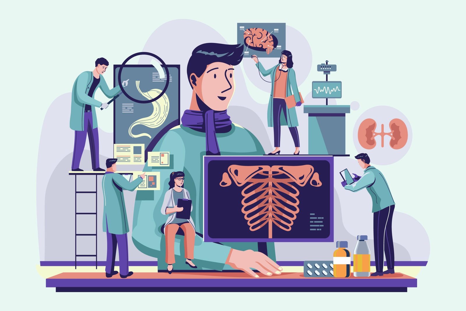
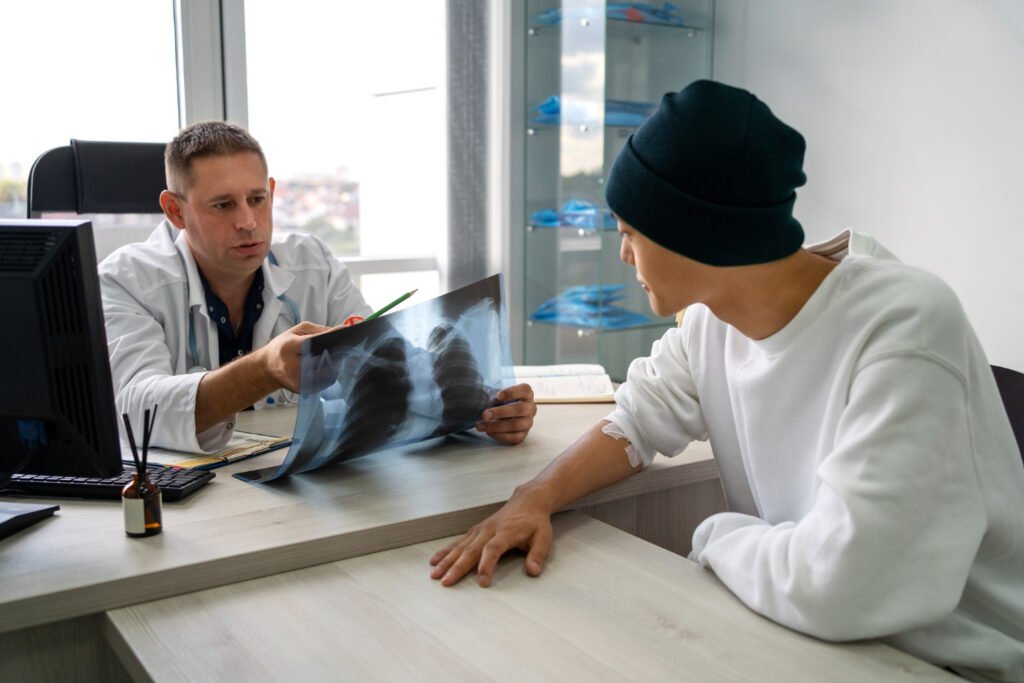
Diagnosis 

The clinical examination is the basis of diagnosis of Graves Disease with the help of laboratory and imaging diagnostic tests.

Clinical Examination:

  • Physical symptoms like an enlarged thyroid gland (goiter), fast heartbeat, tremor and the Graves eye signs (exophthalmos and eye swelling) are examined.
  • Symptoms of hyperthyroidism include weight loss, heat intolerance and nervousness.

Blood Tests:

  • Thyroid hormone levels: TSH is typically very low or not measurable at all because it is inhibited by excess thyroid hormone.
  • Free T4 and Free T3: High levels are a positive evidence of hyperthyroidism.
  • Thyroid antibodies: Thyroid-stimulating immunoglobulins (TSI) or TSH receptor antibodies are a strong indication of Graves Disease.
  • Autoimmune thyroid disease may also be assessed by antibodies to thyroid peroxidase (TPO) and to thyroglobulin.

Radioactive Iodine Uptake (RAIU) Test:

  • Radioactive iodine in small doses is given, and a scan is done to check the absorption of iodine by the thyroid.
  • The diffuse uptake of iodine is normally high in Graves Disease, which can help distinguish it among the causes of hyperthyroidism.

Imaging:

  • Doppler thyroid ultrasound can be done to assess the size, structure, and circulation of the gland.
  • In cases of Graves ophthalmopathy eye imaging (ultrasound, CT scan, MRI) can be utilized to check the eye muscle swelling and orbital involvement.

Additional Tests:

  • In atypical or complex cases, further testing or consultation with endocrinologists may be required.
  • Biopsy of the thyroid is rarely needed.

The combination of these clinical signs, hormone tests, antibody tests, and radioactive iodine uptake has enabled a good diagnosis of Graves Disease and separation from other thyroid conditions.

You may also like to read: Impetigo

 Treatment Options

Graves Disease treatment options are intended to regulate the excessive production of thyroid hormone, alleviate the symptoms, and avoid complications. The major lines of treatment are medication, radioactive iodine therapy and surgery.

Medications

  • Antithyroid Drugs: Methimazole and propylthiouracil (PTU) are widely used to inhibit the production of thyroid hormones. Methimazole is used unless in pregnancy, in the first trimester, PTU is better. Assessing remission often takes 12-18 months, although treatment can require some patients more time. Side effects may include rash, sore joints, liver issues and a rare possibility of low white blood cell count.
  • Beta Blockers: These do not influence thyroid levels but are useful in managing such symptoms as rapid heartbeat, tremor, anxiety and heat intolerance. Widespread beta blockers are propranol and atenol.

Radioactive Iodine Therapy (RAI)

  • Radioactive iodine (I-131) is administered orally, and over weeks or months destroys overactive thyroid cells causing reduced synthesis of thyroid hormones.
  • RAI is a definite therapy that is usually applied when medication fails or in cases of recurrence.
  • It usually leads to hypothyroidism in the future, which needs lifelong replacement of thyroid hormones.
  • It is mostly not used during pregnancy and breastfeeding.
  • It can aggravate or cause thyroid eye disease in patients.

Surgery

  • Thyroidectomy: This is surgical removal of the entire or part of thyroid gland.
  • Surgery can be suggested when the patient is unable to bear antithyroid medicines, has large goiters that lead to compression and in those with severe eye disease.
  • Patients should be put on lifelong thyroid hormone replacement therapy after surgery.
  • Damage to the parathyroid glands (calcium levels) and injury to the vocal cord nerve are the risks, but they are not common in the hands of skilled surgeons.

Choosing Treatment

  • Treatment is determined by patient factors such as age, severity, pregnancy, eye involvement, and patient preference.
  • Others can begin with medications to stabilize, followed by final treatments such as RAI or surgery.
  • Follow-up is necessary to make changes to therapy and complications management.

Lifestyle and Self-Care Tips

Lifestyle and self-care is a supportive aspects to treat Graves Disease along with medical treatment. Major guidelines to enhance the well-being and symptom control are:

Stress Management

  • Use relaxation methods like deep breathing, meditation, journaling or your pleasure causes.
  • Learn to say no and manage time to reduce stress, do not overload oneself.
  • Mood wellness promotes immune homeostasis and may assist with symptoms.

Healthy Sleep and Rest

  • Make getting adequate and regular sleep a priority in order to fight fatigue and enhance general health.
  • Do not stay up late or wear oneself out.

Diet and Nutrition

  • Consume a healthy diet full of antioxidants in fresh vegetables and fruits to alleviate inflammation.
  • Take a sufficient amount of calcium and vitamin D to maintain bone strength and prevent osteoporosis.
  • Add protein-rich foods such as fish, poultry, eggs, and plant proteins to build muscle.
  • Restrict or watch the intake of iodine as recommended by a medical professional, as too much of iodine may increase symptoms.
  • Limit caffeine-based products (coffee, tea, chocolate, soft drinks), which can aggravate palpitations and anxiety.
  • Avoid foods that are highly processed, include sugars, and solid fats; eat whole grains, olive oil, and baked or broiled items.

Exercise

  • Do light exercise every day.
  • Do not overexert, particularly when the heart rate is high.

Social Support and Advocacy

  • Get family, friend, or support group support.
  • Educate yourself on the disease and promote proper care.

Other Tips

  • Adhere to medical regimens.
  • Smoking should be avoided because it aggravates Graves eye disease.
  • Check with health practitioners on thyroid levels and symptoms regularly.

The lifestyle changes can alleviate symptoms, enhance quality of life, and complement the medical therapy of Graves Disease.

Final Thoughts

Graves disease is a complicated autoimmune disorder of the thyroid and may cause severe health problems when unchecked. Majority with early diagnosis, appropriate treatment, and lifestyle changes can easily control their symptoms and lead a good life. Monitoring and collaborating with medical professionals is the principle of managing Graves disease and avoiding chronic problems.

What Is Impetigo? A Complete Guide to This Contagious Skin Infection

Dr. Ahmad Shahzad
Founder | Lyallpur Diabetes Foundation
Consultant Diabetologist | Educator | Advocate for Preventive Care

Impetigo is an infectious and widespread skin disease that occurs both in children and in adults, but in young children, it occurs most frequently. It starts with red sores/blisters and within a short time they become widespread and develop honey colored crusts often on the nose, mouth, hands or even on the open skin. Although impetigo might appear frightening, it is most often mild and is responsive to treatment at an early stage. It is important to know the causes, symptoms, and prevention measures to prevent the spread of the infection and guarantee a rapid cure.

What Is Impetigo?

Impetigo is a widespread, highly contagious bacterial dermatological infection of the epidermis. It occurs most commonly as itchy or painful sores and blisters that burst and leave yellowish or honey-colored crusts particularly on the face, arms, and legs. It is very common in young children between 2 to 5 years old but maybe at any age. Staphylococcus aureus or Streptococcus pyogenes bacteria cause the infection. Transmission of impetigo occurs readily by contact, particularly when there is a large crowd or the skin is torn by wounds, insect bites, or eczema. Therapy typically involves topical or oral antibiotics, and recovery tends to be without scarring. Most common forms include non-bullous impetigo (with tiny blisters and crusting), bullous impetigo (large blisters filled with fluid), and ecthyma (ulcers and severe).

Types of Impetigo

The main types of impetigo are:

  • The most common is non-bullous impetigo (also known as crusted impetigo). It begins with small blisters which burst rapidly, producing red wet spots of skin that form a yellowish-brown or honey-like crust. It normally appears near the mouth, forearms, nose, and hands. Staphylococcus aureus or group A streptococcus bacteria is the cause of most cases.
  • Bullous impetigo results in greater fluid blisters that can be left intact. These blisters tend to be transparent or opaque and are common to the trunk, arms, and legs, particularly among infants and very young children. It is only caused by Staphylococcus aureus, which produces a toxin that causes the upper layer of skin to separate.
  • A more serious and infrequent type is called ecthyma. It produces painful, pus-filled ulcers that go deeper into the skin under the surface, encircled by yellow or brown-black crusts. Ecthyma may leave scars and is a common complication of untreated impetigo.

Thus, impetigo includes these distinct forms: non-bullous (crusted), bullous (blistering), and ecthyma (ulcerative).

Causes and Risk Factors

Bacteria, particularly Staphylococcus aureus and group A Streptococcus (Streptococcus pyogenes) are the most common cause of impetigo. These bacteria enter the skin via breaks like cuts, scratches, insect bites, or any other trauma of the skin. Transmission occurs by direct contact with the sores of an infected individual or by indirect contact through contaminated towels, clothing, or bedding.

Key risk factors that increase the likelihood of developing impetigo include:

  • Children between 2 and 5 years old because their immune system is not fully established.
  • Residing in humid climates, which promotes the growth of bacteria.
  • Open wounds or any other skin disorders like eczema, dermatitis, scabies, insect bites or infectious dermatitis.
  • Bacterial spread is enabled by close living or playing conditions, like in schools, daycare centers or crowded areas.
  • Poor hygiene and lack of hand, body or face washing.
  • Weakened immune system or underlying health problems, including diabetes, HIV / AIDS, cancer therapies, liver or kidney issues, or injecting drugs.

Secondary impetigo may occur when the bacteria infect an already existing skin wound. Trauma, burns, scratching, and any other type of skin injury act as entrances to bacteria. Impetigo can cause complications without treatment, and it is extremely contagious when there are blisters or sores.

Symptoms of Impetigo

Impetigo symptoms vary slightly depending on the type but generally include:

  • Blisters or reddened sores, usually of the area around the mouth and nose, but they may occur anywhere on the body, particularly on the face, arms, and legs.
  • The sores can begin as tiny blisters, which can be itchy or painful and then burst, releasing fluid or pus.
  • These ruptured blisters then develop an archetypal yellowish or honey-colored scab or crust.
  • Scratching or touching may cause the spread of the rash to other parts of the body or nearby.
  • The clusters of blisters may become larger and larger.
  • Under the crust the affected skin can be moist and reddish.
  • Bigger fluid-filled blisters are present in bullous impetigo, which break, leaving crusted sores, which tend to be more painful and swollen.
  • A more severe form is called ecthyma and results in painful sores that go deeper into the skin with thick crusts and can leave behind scars.
  • In more serious cases, some individuals can have swollen lymph nodes in the region or a mild fever.
  • Itching and soreness are frequent but mild as a rule.

You may also like to read: Rhinovirus

How Impetigo Spreads

The transmission of impetigo occurs primarily during direct physical contact with an infected individual, particularly the sores, the blisters or the crusted lesions. It might also propagate indirectly by contact with substances contaminated with the bacteria, such as towels, clothing, bedding, toys or sports equipment. The bacteria causing it- Staphylococcus aureus and Streptococcus pyogenes can survive on these surfaces to infect others.

The other spread factors are those in close contact such as schools, daycare centers, gyms, and sports teams, in particular wrestlers and athletes with a lot of skin contact. Cuts, scrapes, insect bites, or skin diseases like eczema weaken the skin’s defenses by allowing the entry of bacteria.

Impetigo bacteria might also be transmitted through respiratory droplets through coughing or sneezing, and contact with the mucous membranes or skin.

Infection is contagious until the sores are completely cured and the crusts dry out. Hygiene, not sharing personal objects, covering sores, washing hands frequently, and disinfecting surfaces are useful in preventing transmission.

Diagnosis 

The diagnosis of impetigo is mostly based on a history and physical examination. Impetigo is normally diagnosed by a healthcare provider looking at the typical look of the sores, blisters, and crusts on the skin-honey-colored crusts in non-bullous impetigo or bigger, fluid-filled blisters in bullous impetigo. The location and nature of lesions are important diagnostic indicators.

Additional diagnostic methods include:

  • Bacterial culture or swab in the pustules, blisters or crusts may be obtained when there is a suspicion of antibiotic resistance (e.g., MRSA), or when the patient does not respond to conventional therapy. This assists in determining the causal bacteria and in informing the selection of antibiotics.
  • A rare but possible use of skin biopsy is with refractory or atypical cases.
  • Impetigo is not regularly tested with serologic tests of streptococcal antibodies but could be helpful in the event of post-streptococcal complications.
  • Lab testing is not typically required as impetigo diagnosis can be determined through clinical examination.

Differential diagnosis includes other blistering or crusting skin conditions like eczema, herpes simplex, insect bites, contact dermatitis, and fungal infections, so careful skin examination is important.

Treatment Options 

The treatment options for impetigo include both topical and systemic antibiotics depending on the severity and extent of the infection.

  • Topical antibiotics are the first option to localized, simple non-bullous impetigo. Topical agents are typically mupirocin, retapamulin, and fusidic acid (not in every country). The crusts must be washed with soap and water before using them to ensure maximum medication absorption.
  • In the case of bullous impetigo, non-bullous impetigo with over five lesions, deep tissue involvement, or systemic (fever, lymphadenopathy) signs, oral antibiotics are prescribed. Typical oral antibiotics are cephalexin, dicloxacillin or amoxicillin-clavulanate. In case culture demonstrates the presence of methicillin-resistant Staphylococcus aureus (MRSA), clindamycin, doxycycline, or trimethoprim-sulfamethoxazole can be used in place of it.
  • General hygiene should also be adhered to: it is important to keep lesions clean, cover sores with bandages to prevent transmission, wash hands regularly, wash clothes and linens, and not to interact with other people during contagious periods.
  • The average length of treatment is 5-10 days, and the patient must continue the full course despite symptom improvement to avoid resistance and relapse.
  • In recurrent or chronic cases, bacteria carried in the nose is tested and treated with mupirocin nasal ointment.
  • In the case of small, minor diseases, over-the-counter antibiotic creams like bacitracin can be tried, although prescription antibiotics usually work better.

Final Thoughts

Impetigo can be a very contagious skin infection that can be effectively treated with proper awareness, early diagnosis and prompt treatment. The right care clears most of cases in a few weeks with no long-term consequences. The best way to control its spread is by practicing good hygiene, not sharing personal items, and seeking medical assistance whenever symptoms emerge. Knowing the causes, symptoms, and prevention, children and adults can remain safe and get through impetigo soon.

What Is Rhinovirus? Everything You Need to Know About Cold Infections

Dr. Ahmad Shahzad
Founder | Lyallpur Diabetes Foundation
Consultant Diabetologist | Educator | Advocate for Preventive Care

Rhinovirus is the leading cause of the common cold, affecting millions of people worldwide every year. This virus is a highly contagious virus belonging to the Picornaviridae family with a rapid spread via respiratory droplets, contaminated surfaces, and close contact. Although rhinovirus infections are typically mild, they may be very uncomfortable and even lead to complications, particularly in children, the elderly, and immunocompromised individuals. The rhinovirus, its mechanism, symptoms, and prevention methods are important in ensuring improved health.

What Is Rhinovirus?

The rhinovirus is a human pathogen, a positive-sense, single-stranded RNA virus. It causes the common cold, mild cold, or flu-like symptoms of sore throat, runny nose or stuffy nose, sneezing, cough, and nasal congestion. Nasal epithelial cells in the airway are infected by rhinoviruses, which spread via aerosol, respiratory droplets, direct contact, and contamination of surfaces. Rhinovirus A, B, and C have more than 165 types. Infection with rhinovirus is typically mild and may lead to a more serious disease in infants, the elderly, and the immunocompromised. Rhinovirus infections do not have any vaccines or particular antiviral medications.

How Does Rhinovirus Spread?

The rhinovirus is mainly transmitted by the respiratory droplets expelled when an infected individual coughs or sneezes, talks, or breathes. People around can be infected by breathing in these droplets. Direct contact, including shaking hands or touching contaminated surfaces with nasal secretions of a sick individual and then touching the eyes, nose, or mouth (self-inoculation) is also a method of virus transmission. Rhinovirus is very contagious, and infected surfaces or objects can support the virus over several days. Aerosol transmission is also thought to be an important method of transmission, particularly in indoor settings. Individuals become the most infectious during the initial few days of infection when the viral levels in the nasal secretions are the highest.

Symptoms 

The symptoms of a rhinovirus infection, which is the primary cause of the common cold, typically include:

  • Runny or blocked nose
  • Sneezing
  • Sore throat and ears
  • Coughing
  • Watery eyes
  • Headaches and mild body aches
  • Feeling unwell or fatigued
  • Fever (more common in infants and children)
  • Nasal congestion
  • Mild headache
  • Low-grade fever (temperature over 38°C)
  • In infants and children: fever, otitis media (middle ear infection), and potentially more severe infections like croup, bronchiolitis, or pneumonia
  • Asthma attacks may be triggered in some individuals

The symptoms do not exceed 2 weeks and are typically less than 7 days. Infants, the elderly, and individuals whose immune systems are weakened may be affected worse by the infection. Rhinovirus symptoms begin with a sore throat or throat tickling and then move to nasal symptoms and cough.

Potential Complications

Potential Complications of rhinovirus

Possible complications of rhinovirus infection, especially severe cases or vulnerable populations (infants, the elderly, weakened immune system, and people with chronic respiratory diseases), include:

  • Eardrum or middle ear infection (Otitis media).
  • Sinusitis (inflammation or infection of the sinuses)
  • Attacks of asthma and other reactive airway diseases, leading to more airway inflammation and blockage.
  • More serious breathing diseases, bronchitis and bronchopneumonia.
  • Secondary bacterial pneumonia, particularly common in people with malnutrition, excessive cold, or poor immunity.
  • Severe pneumonia can cause acute respiratory distress syndrome (ARDS) and necessitate intensive care with mechanical ventilation.
  • Multi-organ dysfunction syndrome (MODS) is a rare manifestation of severe infections with rhinoviruses.
  • Very rare cases of central nervous system involvement, such as encephalitis and neurological complications, have been reported.
  • Heightened danger of lower respiratory illness, and possibly hospitalization and prolonged sickness.
  • Deaths among high-risk groups, including immunocompromised transplant beneficiaries and cancer patients, have been reported.

There are no particular antiviral interventions, so treatment is supportive, aimed at relieving symptoms as well as enhancing the immune response.

Diagnosis of Rhinovirus

Rhinovirus infection diagnosis is mainly based on the clinical presentation and symptomatology of the patient. Nevertheless, laboratory testing is employed to confirm and make a differential diagnosis, particularly in severe cases or in immunocompromised patients. Molecular detection by reverse transcription polymerase chain reaction (RT-PCR) is the most reliable and employed diagnostic technique that amplifies the presence of specific RNA sequences of the virus in respiratory specimens (nasopharyngeal or oropharyngeal swabs and nasal aspirates) by replicating the virus gene sequences. RT-PCR is sensitive and specific and can identify rhinovirus in many cases within a day. Conventional virus isolation in cell culture is more time-consuming and has been generally substituted by molecular methods. Less sensitive than RT-PCR are antigen detection tests, which do exist, though. Generally, it is advisable to use molecular methods, which are accurate and fast to detect rhinovirus infection.

Treatment and Management

Treatment and management of rhinovirus infection primarily focus on symptomatic relief because the infection is usually mild and self-limited, with no specific antiviral treatments or vaccines available. Key approaches include:

  • Rest and hydration to give the body an immune and recovery boost.
  • OTC pain relievers like acetaminophen and nonsteroidal anti-inflammatory drugs (NSAIDs) like ibuprofen are used to relieve symptoms including headache, muscle ache, fever, and sore throat.
  • Oral or intranasal nasal decongestants can cause nasal congestion, but an intranasal decongestant such as oxymetazoline should be no longer than 3 days to prevent rebound congestion.
  • Anticholinergic effects on the rhinorrhea (runny nose) of the first-generation antihistamines may result in sedation.
  • Persistent rhinorrhea may be decreased with inhaled ipratropium.
  • Taken within 24 hours of onset, zinc lozenges can shorten the length and intensity of symptoms, including cough and runny nose.
  • Resistance to antibiotics, which do not affect viral diseases.
  • Symptomatic car,e like antihistamines alone, antitussives, expectorants, steam inhalation, and numerous herbal preparations have little or no obvious evidence of effect.
  • There are preventive measures, such as good hygiene to reduce the spread of the virus.
  • Soluble ICAM-1 and 3C protease inhibitors have already been examined experimentally but are not available as of now.

You may also like to read: Charcot Foot

Prevention of Rhinovirus Infections

Prevention of rhinovirus infections primarily involves good hygiene and behavioral measures to reduce virus transmission:

  • One of the best prevention methods is regular and thorough handwashing using soap and water to remove the virus on hands.
  • Washing hands with alcohol based hand sanitizers where there is no soap and water.
  • Do not touch the face, particularly the eyes, nose, and mouth, with contaminated hands to avoid self-inoculation.
  • Coughing and sneezing with a tissue or elbow and disposing of the tissue well.
  • Washing and disinfecting commonly touched surfaces and objects such as doorknobs, toys, and tabletops.
  • To mitigate the transmission of the virus, one should avoid coming into close physical contact with cold symptoms and remain at home when unwell.
  • The transmission of respiratory droplets may also be minimized by wearing face masks in busy or risky locations.
  • Balanced nutrition, hydration, and avoiding smoking are all helpful in maintaining good overall health and supporting immune defense.
  • Zinc supplementation in children could help lessen the occurrence of colds.
  • Other preventive interventions, such as gargling, probiotics, and vaccination (not rhinovirus-specific) have marginal or no definitive value.

Final Thoughts

Overall, rhinovirus is a common but usually mild infection that remains the leading cause of the common cold. Although there is neither a specific cure nor a vaccine, with proper hygiene, healthy lifestyle, and management of the symptoms, its impact can be significantly diminished. Knowing the transmission mechanism of rhinovirus and simple precaution measures will help people to keep themselves and others safe, particularly those who may be predisposed.