Diabetes Pakistan Metabolic Syndrome Date: 1st - 2nd November, 2024 Venue: Serena Hotel, Faisalabad
For Emergencies: +92 41-2694037

The Impact of Diabetes on Bone Health

Dr. Ahmad Shahzad
Founder | Lyallpur Diabetes Foundation
Consultant Diabetologist | Educator | Advocate for Preventive Care

As we know, diabetes can have an impact on blood sugar and general metabolic wellbeing, but that is just the tip of the iceberg. Bone health is a field that is not given much attention. As per the research, individuals with diabetes have a greater risk of fractures, low bone density, and low bone strength. It is important to understand the impact of diabetes on the skeletal system to prevent this condition, detect it early and improve the long-term health outcomes.

Understanding the Link Between Diabetes and Bone Health

Diabetes causes serious complications in bone health, bone density, and bone quality, predisposing individuals to fracture. Type 1 diabetes (T1DM) and type 2 diabetes (T2DM) both cause bone problems, albeit in slightly different ways.

Impact on Bone Density and Structure

·       T1DM is typically linked to reduced bone mineral density (BMD), which disables bone formation. This is attributed to hyperglycemia leading to accumulation of advanced glycation end products (AGEs) which destroy bone cells, disrupt the activity of osteoblasts (bone-building cells) and augment bone resorption by osteoclasts. T1DM also depletes growth factors like insulin-like growth factor-1 (IGF-1), which also inhibits bone growth and bone remodeling.

·       T2DM, ironically, is commonly characterized by normal or even better BMD but worse bone quality. The prolonged hyperglycemia and related metabolic alterations worsen the organic structure and biomechanical characteristics of the bone, making bones weaker despite increased density. The bone-forming cells are also adversely affected by increased bone marrow fat and inflammation in T2DM.

Increased Fracture Risk and Healing Complications

·       T1DM and T2DM patients are more likely than non-diabetics to suffer a fracture. This is not necessarily attributed to structural integrity of the bone but also due to complications of diabetes like neuropathy and muscle weakness, eye issues and frequent falls which increase the risk of fracture.

·       The recovery of fractured bones in diabetics may be delayed due to impaired bone cell activity, reduced progenitor cells and poor vascularization of fracture locations. Long-term hyperglycemia may have permanent adverse effects on bone cell activity, which impairs proper repair.

Clinical and Treatment Considerations

  • Prevent or reduce diabetic bone disease through good glycemic control. Certain antidiabetic drugs (e.g., metformin) are potentially better since they have fewer adverse effects on bone health, whereas other drugs (such as thiazolidinediones) can increase bone loss.
  • Therapies focused on the enhancement of bone density and quality, including anti-resorptive medications (e.g., alendronate), bone-forming (e.g., intermittent PTH in T1DM), and vitamin K2 and vitamin D3 supplementation in T2DM, have potential in the treatment of bone complications in diabetes.
  • As well as acting to regulate blood sugar, insulin therapy has direct positive effects on bone formation by stimulating osteoblastic activity.

Mechanisms: How Diabetes Affects Bones

Diabetes affect bone health through several interconnected mechanisms centered around hyperglycemia, insulin deficiency or resistance, and inflammation, which alter bone cell function, bone matrix quality, and bone remodeling processes.

Key Mechanisms

  • Hyperglycemia and Advanced Glycation End Products (AGEs): Chronic high blood sugar causes the development of AGEs that build up in bone collagen. This deposition makes the collagen matrix weaker and less durable, which reduces the mechanical strength and quality of bone. AGEs also induce bone progenitor cell apoptosis (death), osteoblast differentiation and function, and osteoclastic activity, resulting in elevated bone resorption.
  • Impaired Osteoblast Function and Reduced Bone Formation: In type 1 diabetes (T1DM), there is a lack of insulin and growth factors such as insulin-like growth factor 1 (IGF-1) that inhibits the differentiation of mesenchymal stem cells (MSC) to osteoblasts. New bones are formed by osteoblasts, and thus their dysfunction reduces bone formation and bone mineral density (BMD). Repeated hyperglycemia suppresses important genes and proteins in osteoblast maturation and activity as well.
  • Disturbed Bone Remodeling Dynamics: Diabetes causes disequilibrium in bone remodeling. The AGEs and the inflammatory cytokines enhance the activity of osteoclasts (bone resorption cells) and reduce the activity of osteoblasts, impairing normal bone turnover. This leads to poorer bone structure and fragility even with normal or even higher BMD particularly in diabetes type 2 (T2DM).
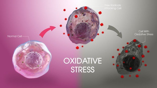
  • Inflammation and Oxidative Stress: High blood sugar increases oxidative stress and proinflammatory pathways (e.g., NF-kB) and releases cytokines, such as TNF-a and IL-6. Such cytokines also suppress osteogenic differentiation of bone marrow stem cells and bone repair. Ironic inflammation also leads to bone quality and healing capacity.
  • Bone Marrow Adiposity and Cellular Changes: In T2DM, bone marrow fat accretion leads to release of free fatty acids and pro-inflammatory cytokines, which worsen insulin resistance and suppress osteoblast activity. Bone marrow population of MSC also reduces which hinders the formation of new bone cells and restoration of broken bones.
  • Vascular and Healing Losses Diabetes-induced loss of MSC viability and function also impairs vascularization of fracture sites, resulting in delayed bone healing, increased incidence of non-union fracture, and postoperative complications in diabetic patients.

Simply put, diabetes impairs bone health by exerting hyperglycemia-induced biochemical effects, inflammation, and altered stem cell processes that increase the risk of fracture and decrease bone repair potential.

Increased Risk of Fractures in People with Diabetes

Strategies for Protecting Bone Health in Diabetes

Individuals with diabetes, especially type 2 diabetes (T2DM), are at a higher risk of developing bone fractures because of multifactorial etiological factors other than bone mineral density (BMD). This increased risk of fracture is due to several factors:

Risk Factors

  • Bone Quality Impairment: Although many T2DM patients have normal or increased BMD, their bone quality is impaired by the deposition of advanced glycation end products (AGEs), low bone turnover, and alteration of bone microstructure, which impair bone tissue mechanical strength and strength.
  • Duration and Severity of Diabetes: Long diabetes time (particularly 15 years and above) and inadequate glycemic control raise the risk of fractures. Fracture risk is also linked to insulin therapy which often is a sign of severe or advanced disease.
  • Age and Body Mass Index (BMI): Elderly diabetic patients are at risk of getting a fracture. There is a significant increase in the risk of fractures in diabetic patients with a low BMI (<25 kg/m2), and a moderate effect of higher BMI.
  • Diabetes Complications and Hypoglycemia: Diabetes-related conditions, including peripheral neuropathy, muscle weakness, impaired vision, and hypoglycemia (particularly because of drug use) are known to put patients at risk of falls and fractures.
  • Medications: Thiazolidinediones, SGLT2 inhibitors, and insulin are some diabetes therapies that have been associated with an increased risk of fractures either via their impact on bone metabolism or by raising the risk of falls.
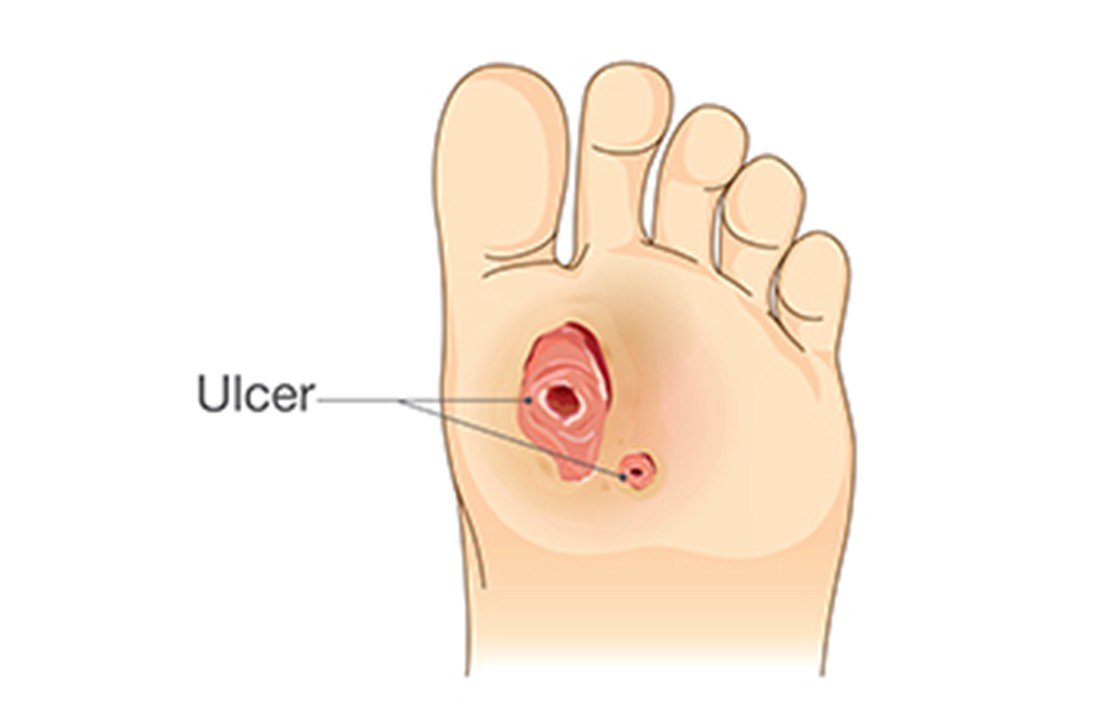
  • T2DM patients experience higher fractures at the hip, vertebrae, and feet, but not always wrists, shoulders, or ankles.

Additional Factors Contributing to Poor Bone Health in Diabetes

Additional factors contributing to poor bone health in people with diabetes go beyond changes in bone mineral density (BMD) and include both skeletal and extra-skeletal influences:

  • Advanced Glycation End Products (AGEs) Accumulation Hyperglycemia increases AGEs formation, changes bone collagen, making bones more rigid, fracture-prone, and brittle. This affects bone quality without BMD.
  • Raised Fall Risk: Diabetes complications like peripheral neuropathy, impaired balance, muscle weakness, vision issues, and hypoglycemic episodes predispose one to fall, significantly contributing to the risk of fractures.
  • Negative Effect of Some Medicines: Some antidiabetic medications (e.g. thiazolidinedions) have been shown to increase the risk of fractures by either impairing the formation of bone or enhancing its resorption. There are certain reservations about SGLT2 inhibitors that are mixed. Insulin therapy is anabolic to bone on its own, and usually a sign of advanced diabetes that correlates with bone fragility.
  • Physical Inactivity: Diabetes complications reduce mobility, which slows down mechanical loading of bones and increases bone loss and skeletal weakness.
  • Inflammation and Oxidative Stress: Chronic inflammation caused by diabetes and oxidative stress destroys bone cells, alters the work of osteoblasts and stimulates bone resorption.
  • Finally, The Bone Marrow Composition Changes Type 2 diabetes alters the balance of bone marrow fat content which disrupts normal bone remodeling by preventing osteoblast differentiation and enhancing adipogenesis in bone marrow.
  • Vitamin D Deficiency and Calcium Imbalance: Diabetes may be associated with a lack of vitamin D and poor calcium metabolism to weaken bone mineralization and strength.
  • Comorbid Conditions: Diabetes may be comorbid with many conditions, including kidney disease, autoimmune diseases (e.g., celiac disease, Graves’ disease), hormone imbalance, and eating disorders, all of which may lead to poor bone health.
  • Genetic Factors: Genetic polymorphisms in collagen synthesis genes and vitamin D receptor genes can also be involved in bone fragility in diabetes.

You may also like to read: Connection between Diabetes and Cognitive Decline

Strategies for Protecting Bone Health in Diabetes

Treatment of bone in diabetic individuals is a multifaceted process that involves proper glycemic control, lifestyle changes, nutritional interventions, drug-specific selection and, in some cases, bone-specific therapy.

Key Strategies

  • Optimal Glycemic Control: Good blood sugar control minimizes the development of damaging advanced glycation end products (AGEs) in bone tissue and the risk of diabetic complications such as neuropathy and hypoglycemia that elevates the risk of falls and fractures. It is important to prevent hypoglycemic episodes by avoiding frequent ones.
  • Lifestyle and Physical Activity: Resistance and weight bearing exercises (walking, strength training) promote bone density and bone strength. Exercise also enhances muscle tone and stability, which prevent falls. Quitting smoking and alcohol reduction are essential because these two habits undermine bones and predispose them to fractures.
  • Nutritional Support: Optimal consumption of calcium and vitamin D is the key to good bone health. When the dietary intake is inadequate or vitamin D is low, supplements may be required especially in type 1 diabetes and the elderly with type 2 diabetes. Proteins are useful to maintain bones and muscles.
  • Drug prescribing and Reimbursement: Select bone-neutral or bone-positive diabetes agents, including metformin, GLP-1 receptor agonists, and DPP-4 inhibitors. Thiazolidinediones (TZDs) should be avoided and SGLT2 inhibitors should be monitored because they may increase the risk of fractures. Insulin is to be administered with caution to prevent hypoglycemia.
  • Bone-Targeted Therapies: In patients with osteoporosis or with a high risk of fracture, antiresorptive medications such as bisphosphonates (e.g., alendronate) and anabolic therapies such as teriparatide (PTH 134) have demonstrated effectiveness in enhancing bone density and fracture risk. Vitamin K2/D3 combination therapy has been demonstrated to induce bone formation in diabetic bones.
  • Bone Health Check: It is essential to monitor bone mineral density (BMD) and bone turnover, as well as assess fracture risk, to ensure prompt treatment. Improved risk prediction of fractures in diabetes might be achieved by newer imaging methods and customized risk models.
  • Make improvements to Fall Risk and Comorbidities: Fall management: treat diabetic neuropathy, visual issues, and muscle weaknesses to decrease falls. Vitamin D deficiency and other comorbidities of bone health are significant aspects of care that require screening and treatment.

Final Thoughts

Finally, diabetes does not only influence the level of blood sugar but also has an important effect on the strength of bones and general skeletal health. Diabetes demonstrates the need to make proactive care a priority by raising the risk of low bone density, fractures, and slow healing. It can be avoided by keeping blood sugar levels, consuming a balanced diet with bone strengthening nutrients, exercise, and regular examinations. People with diabetes can minimize the complications and help secure a healthier and stronger future with the right preventive measures.

The Connection between Diabetes and Cognitive Decline

Dr. Ahmad Shahzad
Founder | Lyallpur Diabetes Foundation
Consultant Diabetologist | Educator | Advocate for Preventive Care

The effect of diabetes on blood sugar control is well known; however, its effects extend far beyond just physical health. Studies indicate a Connection between Diabetes and Cognitive Decline, attributing the disease to memory loss, diminished attention and even increased dementia susceptibility. This relationship is paramount as it reveals the necessity of controlling diabetes to not only preserve the body but also the health of the brain in the long term.

Understanding Cognitive Decline

Cognitive decline is a progressive loss of mental abilities like memory, reasoning and attention. It is commonly an aging process but may be hastened or aggravated by multiple factors such as illnesses and habits.

Symptoms

Symptoms can vary but often include:

  • Memory loss or trouble recalling recent events.
  • Difficulty finding words or expressing oneself.
  • Getting lost in familiar places.
  • Trouble with concentration and complex tasks.
  • Mood changes such as increased anxiety or irritability.

How Diabetes Affects the Brain

Brain- Diabetes has several serious effects on the brain, including cognitive impairment, memory loss and dementia. Brain dysfunction is caused by both type 1 and type 2 diabetes, but type 2 diabetes is more closely associated with faster brain aging and dementia, such as Alzheimer disease and vascular dementia.

Mechanisms of Brain Impact

  • Diabetes leads to injury of small and large blood vessels in the brain that limits blood flow and oxygen supply to the brain thus affecting the functioning of brain cells leading to stroke and vascular dementia.
  • And high blood sugar (hyperglycemia) and insulin resistance impair brain metabolism. Brain insulin resistance may also change how energy is used and lead to accumulation of harmful proteins (amyloid and tau) that cause Alzheimer disease.
  • There is observed brain atrophy (shrinkage), especially in memory and executive functions such as hippocampus, medial temporal, and frontal lobes in individuals with diabetes.
  • Neuroinflammation and disturbances in the metabolism of neurotransmitters in the brain due to diabetes are other factors that contribute to the neuronal damage and cognitive decline.

Cognitive Effects and Risk

  • Diabetics (type 2) show increased rates of cognitive impairment compared to healthy people; memory, speed of processing, and some executive functions like planning and attention.
  • There are slight variations in brain functions in children with type 1 diabetes that can influence learning and behavior.
  • • The more a person has diabetics and the poorer the blood glucose management, the greater the risk of cognitive impairment and dementia.

Scientific Evidence Linking Diabetes and Cognitive Decline

Scientific Evidence Linking Diabetes and Cognitive Decline

There is strong scientific evidence of the association between diabetes and cognitive impairment, such as, higher risk of dementia and Alzheimer disease. Numerous studies of high quality, systematic reviews and meta-analyses have found this relation based on observational research, neuroimaging and biochemical studies.

Key Evidence and Findings

  • Diabetes worsens cognitive impairment through chronic hyperglycemia, insulin resistance, inflammation, vascular damage, and oxidative stress, which cause brain cell damage and neurodegenerative protein buildup (tau and amyloid-beta).
  • Meta-analyses indicate that individuals with diabetes face an increased risk of getting dementia (59-73 percent higher than non-diabetics) and an almost doubled risk of getting Alzheimer disease.
  • The risk of cognitive loss is increased by diabetes and the occurrence of complications like hypoglycemia.
  • A. Neuroimaging showed accelerated brain atrophy and accelerated cognitive decline in individuals with type 2 diabetes compared to age controls.
  • Mechanistic studies (1) connect neurodegeneration and impaired neurotransmitter balance to diabetes-related brain insulin resistance, mitochondrial dysfunction, and chronic inflammation.
  • Glucose-lowering treatments such as metformin and SGLT-2 inhibitors demonstrate the possibility to decrease dementia risk in diabetic groups, which indicates modifiable aspects in diabetes treatment to reduce cognitive impairment.

Risk Factors and Contributing Elements

Diabetes-related cognitive decline is influenced by multiple risk factors and contributing elements spanning vascular, metabolic, inflammatory, and lifestyle domains.

Major Risk Factors

  • Vascular Disease: Macrovascular (stroke, heart disease) and microvascular (retinopathy, nephropathy) diabetes complicates are significant in terms of cognitive impairment. These risks are worsened by hypertension and dyslipidemia.
  • Hyperglycemia: Persistent elevated blood sugar and glucose levels cause neuronal oxidative stress, mitochondrial dysfunction, and the generation of toxic AGEs, enabling damaging brain inflammation.
  • Hyperinsulinemia and Insulin Resistance: Early insulin resistance and elevated insulin levels are associated with worse cognition and risk of Alzheimer, in part because of impaired brain insulin signaling.
  • Neuroinflammation: The diabetic brain is persistently inflamed with increased levels of inflammatory cytokines and microglial activation leading to neuronal damage resulting in the exacerbation of cognitive impairment.
  • Period and Level of Diabetes: Longer diabetes and high HbA1c decrease the cognitive impairment potential and severity.
  • Other Factors: It is also increased by age (greater than 60 years), female sex, low education level, stroke or depressive history, obesity, smoking and lack of exercise.

Additional Considerations

  • The abnormalities in the brain metabolic markers and neurotransmitters of memory and cognitive processing are usually identified in diabetic patients.
  • Mild cognitive impairment (MCI) is prevalent in older adults with type 2 diabetes, which usually evolves into dementia unless treated.
  • Certain antidiabetic medications such as metformin can protect cognition, but the evidence is inconclusive and needs to be examined further.

You may also like to read: Role of Oxidative Stress in Diabetic Complications

Prevention and Management Strategies

Cognitive decline in diabetes is prevented and managed by maximizing metabolic control, vascular risk factors, and healthy lifestyle behaviors.

Glycemic Control

·       Antidiuretic drugs like metformin and SGLT-2 blockers have demonstrated some neuroprotective effects by risk-reducing dementia and enhancing cognitive performance.

·       Good glucose control is paramount but excessive tight control resulting in a higher risk of hypoglycemia can exacerbate cognition. HbA1c targets are to be individualized depending on cognitive status and age to prevent hypoglycemia.

Cardiovascular and Vascular Risk Management

·       Regular aerobic physical activity is associated with enhanced cardiovascular health, cerebral blood flow, neurotrophic, and inflammation, which in totality safeguard cognition.

·       Management of hypertension, dyslipidemia and other cardiovascular risk factors minimize cerebrovascular loss which causes impairment in cognition.

Lifestyle Interventions

·       Vascular and metabolic health can be supported by a healthy diet, including the Mediterranean diet.

·       Physical activity is one of the most supported methods of preventing age-related cognitive decline and dementia.

·       Cognitive stimulation and socialization are suggested as effective in keeping the brain functioning.

Additional Strategies

  • Prevention of hypoglycemia is vital as it is linked to progressive cognitive impairment and a higher risk of severe events among the cognitively impaired diabetics.
  • Cognitive health is further supported by monitoring and the management of comorbidities such as depression and obesity.
  • Studies of new treatments of brain insulin resistance, inflammation, and amyloid deposition are in progress.

Final Thoughts

Diabetes is also associated with mental degradation, where both biological and lifestyle determinants contribute to the phenomenon. The risks are severe, but the brain can be preserved with proper blood sugar management, healthy lifestyle, and frequent cognitive monitoring. With some proactive measures, people with diabetes can mitigate the risk and help to maintain their physical and psychological health in the long term.

The Role of Oxidative Stress in Diabetic Complications

Dr. Ahmad Shahzad
Founder | Lyallpur Diabetes Foundation
Consultant Diabetologist | Educator | Advocate for Preventive Care

Diabetes is a complicated metabolic condition that could cause severe health problems besides high blood sugar. Oxidative stress is one of the major contributors to these complications, as this is a condition of imbalance in the body between free radicals and antioxidants. It is also proven that oxidative stress contributes significantly to the destruction of blood vessels, nerves, kidneys and other organs and thus is directly connected to diabetic complications. Understanding the role of oxidative stress in diabetic complications is essential for developing better strategies to prevent, manage, and treat long-term effects of the disease.

What is Oxidative Stress?

Oxidative stress refers to a situation in which the formation and accumulation of reactive oxygen species (ROS), or free radicals, and the capacity to eliminate the reactive species or the damage caused by them is imbalanced. The result of this imbalance is cell and tissue damage since the surplus free radicals can damage vital structures in cells like proteins, lipids, and DNA. Although ROS are necessary in normal cellular activities such as signaling, immune defense, high levels of ROS because of environmental factors or metabolic imbalance led to oxidative stress, which causes aging and many diseases, including cancer, diabetes, cardiovascular and neurodegeneration diseases.

This imbalance arises when the free radicals exceed the antioxidant capacity of antioxidants, the molecules that could stabilize free radicals by donating electrons. Pollution, UV radiation, smoking and some drugs are some of the factors that can initiate oxidative stress. Enzymatic antioxidants like superoxide dismutase, catalase and glutathione peroxidase are used by the body to defend itself, yet, when these systems become overburdened, cell damage occurs.

Oxidative Stress and Diabetes: The Connection

Diabetes is closely related to oxidative stress in a complex way where oxidative stress is essential in pathogenesis, progression, and complications of diabetes. Prolonged high blood glucose (hyperglycemia) and mitochondrial dysfunction in diabetes, especially in type 2 diabetes, cause excess production of reactive oxygen species (ROS). Such surges in ROS exacerbate oxidative stress, which harms pancreatic beta cells that produce insulin, disabling their activity and leading to cell death, decreasing insulin production. Several other causes also lead to insulin resistance, including oxidative stress, when the cells of the body cannot react to insulin, further disrupting the glucose regulation.

Besides, oxidative stress contributes significantly to diabetic complications involving various organs. It harms the vascular system by fostering inflammation and endothelial dysfunction resulting in microvascular complications such as diabetic retinopathy (eye destruction), nephropathy (kidney destruction) and neuropathy (nerve destruction). It also leads to macrovascular problems, which are a risk factor of developing cardiovascular diseases in diabetics. The permanent oxidative injury may perpetuate a deleterious hyperglycemic memory, continuing tissue dysfunction despite normalization of blood glucose.

Antioxidant therapies, lifestyle activities, including diet and exercise, as well as careful blood sugar control, are strategies to counter oxidative stress in diabetes. Nevertheless, the mechanisms remain unclear, and more studies are required to maximize the therapies that address oxidative stress to prevent and manage diabetic complications.

Mechanisms Behind Oxidative Stress in Diabetes

Mechanisms Behind Oxidative Stress in Diabetes

Oxidative stress mechanisms in diabetes entail a series of interacting events that cause an overproduction of reactive oxygen species (ROS) and dysfunction of antioxidant responses and play a direct role in the pathogenesis and complications of diabetes.

Sources of ROS in Diabetes:

Mitochondria during cellular respiration are the major source of oxidative stress in diabetes. Hyperglycemia (high blood sugar) leads to excess glucose metabolism via glycolysis and Krebs cycle leading to high levels of electron donors (NADH and FADH2) overloading the electron transport chain. This leads to loss of electrons to oxygen to form superoxide radicals (O2- -) and other ROS like hydrogen peroxide (H2O2) and hydroxyl radicals (- OH). Moreover, there is hyperactivity of enzymatic systems such as NADPH oxidase, which increases the production of ROS in endothelial cells, smooth muscle cells, and fibroblasts especially when hyperglycemia occurs.

Impaired Antioxidant Defense:

Diabetes is known to break the equilibrium by reducing the activity of protective antioxidant enzymes such as superoxide dismutase (SOD), catalase and glutathione peroxidase (GSH-Px). Decreased antioxidants result in accumulating ROS with destruction of cells.

Molecular Pathways Amplifying Oxidative Stress:

Protein Glycation and lipid peroxidation: Non-enzymatic glycation of proteins and lipid peroxidation leads to the production of free radicals and advanced glycation end products (AGEs), which further enhance the production of ROS and inflammation.

  • Polyol Pathway Activation: Aldose reductase converts excess glucose into sorbitol and depletes NADPH and weakens the ability to regenerate antioxidants.
  • Protein Kinase C (PKC) Activation: Hyperglycemia triggers PKC, which promotes NADPH oxidase, elevates inflammatory cytokines and modulates gene expression of cell damage and vascular dysfunction.
  • Hexosamine Pathway: Enhanced activity of this pathway alters transcription factors by O-linked glycosylation and impacts diabetic complication related genes.
  • Chronic Inflammation: ROS stimulate inflammatory transcription factors (e.g., NF-kB) which leads to driving cytokine production and maintaining a vicious cycle of ROS production.

Impact on Insulin and Beta Cells:

The low antioxidant capacity of ROS leads to insulin secretion and beta cell apoptosis as these molecules cause oxidative damage to pancreatic beta cells. Furthermore, oxidative stress causes insulin resistance by disrupting the insulin signaling that occurs in insulin metabolic dysfunction that is typical of type 2 diabetes.

Contribution to Diabetic Complications

Oxidative stress injures endothelial cells, resulting in vascular dysfunction, microvascular (retinopathy, nephropathy, neuropathy) and macrovascular (cardiovascular disease) complications.

Oxidative Stress in Diabetic Complications

Oxidative stress has been at the center of the development and progress of diabetic complications, micro and macro vascular. Chronic elevated blood sugar in diabetes leads to mitochondrial overproduction of reactive oxygen species (ROS) in endothelial cells and other tissues, resulting in several harmful effects that cause diabetic complications.

Key pathways activated by oxidative stress in diabetes complications include:

  1. Polyol Pathway Flux: Surplus glucose is metabolized to sorbitol by aldose reductase, depleting NADPH, decreasing antioxidant regeneration, and predisposing cells to oxidative damage.
  2. Advanced Glycation End-Products (AGEs): High glucose enhances the development of AGEs which bind to their receptors (RAGE), leading to the production of ROS, inflammation and tissue damage observed in diabetic nephropathy, retinopathy and neuropathy.
  3. Activation of Protein Kinase C (PKC): ACTH hyperglycemia-activated PKC isoforms upregulate ROS, inflammation, vascular permeability, and abnormal blood flow, which add to vascular complications.
  4. Hexosamine Pathway: Overflow of glucose flux causes changes in transcription factors that control fibrosis and inflammatory genes, exacerbating tissue injury.
  5. Direct Enzyme Inactivation: ROS directly inactivates protective against atherosclerosis, including endothelial nitric oxide synthase (eNOS) and prostacyclin synthase, disrupting vascular activity.

Activation of transcription factors such as NF-KB through oxidative stress results in a vicious cycle of cytokine release and immune cell recruitment that exacerbates vascular and tissue injury.

Most heavily impacted organs are kidneys (diabetic nephropathy), eyes (retinopathy), nerves (neuropathy), and the cardiovascular system, which cause greater risk of kidney failure, blindness, neuropathic pain, and heart disease. Epigenetic changes which persist pro-inflammatory gene activity in the face of controlled blood sugar, or so-called metabolic memory, are also caused by oxidative stress.

Superoxide dismutase, catalase, and glutathione peroxidase are the antioxidant defenses that are also impaired in diabetes, thus worsening oxidative damage.

You may also like to read: Importance of Foot Care in Preventing Diabetic Foot Ulcers

Role in Beta-Cell Dysfunction and Insulin Resistance

There are multiple ways that oxidative stress contributes to beta-cell dysfunction and insulin resistance in diabetes:

Beta-Cell Dysfunction

  • Susceptibility: Pancreatic beta cells possess low intrinsic antioxidant capacity and are therefore extremely susceptible to oxidative stresses produced by overproduction of reactive oxygen species (ROS) under hyperglycemic and inflammatory conditions.
  • Damage and Death: Overproduction of ROS results in mitochondrial dysfunction and endoplasmic reticulum (ER) stress in beta cells, leading to cell apoptosis and insulin secretion.
  • Loss of Identity: Oxidative stress results in loss of important beta-cell maturity transcription factors like MAFA and PDX1 to cause beta-cell dedifferentiation and decreased insulin synthesis.
  • Inflammation: ROS activate immune systems through pro-inflammatory cytokines (e.g., IFN-7, IL-1 1, TNF-7) that contribute to additional beta cell death, particularly in diabetes type 1.
  • Antioxidant Downregulation: Downregulation of endogenous antioxidants such as superoxide dismutase (SOD2) enhances oxidative beta cell injury.
  • Protective Pathways: FOXO1 to the nucleus is a defense to oxidative stress that can be overwhelmed in chronic conditions.

Insulin Resistance

·       Disruption of the Insulin Signaling: ROS trigger serine/threonine kinases including JNK, p38 MAPK, and IKK that phosphorylate insulin receptor substrates (IRS) on serine residues and disrupt their function in relaying insulin signals in cells like muscle and adipose tissue.

·       PI3K Disruption: Oxidative stress interferes with translocation and activation of PI3K, an insulin signaler, facilitating insulin resistance.

·       PI3K Disruption: Oxidative stress interferes with translocation and activation of PI3K, an insulin signaler, facilitating insulin resistance.

·       Inflammatory Pathways: ROS stimulate inflammatory pathways (NF-KB), prolonging the systemic inflammation thereby worsening further insulin insensitivity.

·       Redox Imbalance: The imbalance between the production of ROS and antioxidant defense is one of the causes of disturbed metabolism and desensitization of insulin receptors in peripheral tissues.

Strategies to Reduce Oxidative Stress in Diabetes

Oxidative stress reduction strategies in diabetes involve a combination of lifestyle change, antioxidant therapy, and appropriate management of the disease to regain redox balance and alleviate cellular injury:

Lifestyle Modifications

  • Diet: The Mediterranean diet is rich in natural antioxidants in fruits, vegetables, whole grains, olive oil, nuts, and fish, and has been shown to be effective in improving antioxidant enzyme activity and reducing oxidative stress markers. The activation of NRF2 pathway by key phytochemicals upregulates the protective antioxidant genes such as glutathione peroxidase and superoxide dismutase. Low-calorie diets decrease the variability of blood glucose and the production of reactive oxygen species (ROS).
  • Exercise: Mediate aerobic exercise (e.g., walking, cycling) enhances mitochondrial activity and increases endogenous antioxidant enzymes, alleviating oxidative injury in diabetic individuals. Resistance training and high-intensity interval training (HIIT) also have potential in improving antioxidant defenses and glycemic control, but individual health status and age can vary in response. Moderate workouts will avoid the overload of ROS that may exacerbate stress.
  • Stress Management and Sleep: Mindfulness, yoga, and sufficient sleep, lower systemic inflammation and cortisol which otherwise contribute to increased oxidative stress and insulin resistance.

Antioxidant Therapies

  • Supplementation: Alpha-lipoic acid, coenzyme Q10, glutathione and vitamins C and E have also been studied to restore insulin sensitivity and prevent oxidative damage, although the clinical benefit is inconsistent.
  • Targeted Therapeutics: New therapeutic strategies target mitochondria-targeted antioxidants and activators of transcription factors such as NRF2 that control antioxidant defenses. It is also being investigated to inhibit NADPH oxidase (NOX) enzymes, which are key sources of ROS.
  • Nanotechnology and Microbiome: new directions involve nanocarrier-delivered antioxidants to pancreatic islands or atherosclerotic foci, or the regulation of the gut microbiota to ease overall oxidative stress in the body.

Effective Diabetes Management

  • Blood Sugar Control: Tight glycemic control by diet, medication and monitoring mitigate excessive ROS generation in response to hyperglycemia and oxidative stress.
  • Medications: some antidiabetic drugs such as metformin and GLP-1 receptor agonists are antioxidants that minimize oxidative stress beyond the effect of glucose.

Final Thoughts

Summing up, oxidative stress plays a key role in most diabetic complications, including nerve and kidney damage and cardiovascular disease. It impairs the long-term outcomes of diabetes by destabilizing the cellular balance and triggering inflammation. Addressing the role of oxidative stress in diabetic complications through lifestyle changes, antioxidant support, and targeted therapies offers a promising path to protect patients’ health and improve quality of life.

The Importance of Foot Care in Preventing Diabetic Foot Ulcers

Dr. Ahmad Shahzad
Founder | Lyallpur Diabetes Foundation
Consultant Diabetologist | Educator | Advocate for Preventive Care

Having diabetes does not only entail checking blood sugar but also taking care of the feet. Foot ulcers are among the worst complications diabetics can get, as they may become infected and result in amputation without treatment. The positive is that this risk can be significantly decreased with proper foot care. Daily foot checks and healthy habits can help people with diabetes to keep their feet safe and enjoy a higher quality of life.

Understanding Diabetic Foot Ulcers

A diabetic foot ulcer is an exposed wound or sore on a foot whose underlying cause is diabetes. It occurs because of the combination of multiple factors such as peripheral neuropathy (damage to nerves), poor circulation (ischemia), foot defects and recurring pressure or trauma. Neuropathy decreases sensation, i.e. injury or pressure, which leads to the ulcer not being noticed and untreated. Low circulation also worsens recovery and risks infection. They are severe complications of diabetes and may result in infection, hospitalization, and amputation unless timely and adequately treated.

Why Foot Care Matters for Diabetics

Foot care is very important to individuals with diabetes since diabetes can lead to nerve damage (neuropathy) and poor circulation, both of which increase the likelihood of foot injuries not being noticed and not healing. Damage of nerves lowers the sensation of pain, heat, or cold, thus simple cuts, blisters, or sores may turn into severe infections before they are noticed. Healing and the ability to combat infection are further affected by poor blood flow. Consequently, ulcers, infections, and even amputations may result when diabetic foot wounds are not properly tackled.

Good foot care prevents these complications by promoting early detection of injuries through daily inspection, skin hygiene and moisture, foot protection through footwear and socks, and frequent check-ups. Socking and shoing feet prevent unseen trauma injuries even at home. Better foot health is also facilitated by management of blood sugar and general control of diabetes. Various studies indicate that active foot care and early interventions decrease the chances of severe infections and amputations. Foot care will therefore be critical component of diabetes self-management to sustain mobility and quality of life.

Essential Foot Care Practices to Prevent Ulcers

Essential Foot Care Practices to Prevent Ulcers

Essential foot care practices to prevent diabetic foot ulcers include the following key measures:

  • Daily Foot Inspection: Inspect both feet daily to observe any cuts, blisters, redness, swelling or any evidence of injury or infection. Look at the soles with a mirror. Early diagnosis aids in the treatment of problems before they aggravate.
  • Foot Hygiene: Wash feet with lukewarm water daily, dry them completely between toes, and keep skin moist to avoid cracks. Do not keep feet long soaked or rub them with harsh chemicals.
  • Dress Shoes: Use well-fitting shoes that are not tight or loose and high-heeled shoes. Wear clean and dry socks in place of tight bands and never walk barefoot to avoid injury.
  • Frequent Nail Maintenance: Clip toenails in a straight line and do not dig into corners to prevent ingrowth and infection of nails.
  • Pre-ulcerative Signs: Calluses, blisters, fissures, fungal infection, ingrown nails should be treated promptly and with proper care or professional assistance.
  • Pressure Offloading: Special footwear, insoles or orthotics are used to alleviate pressure in specific areas that may have foot deformities or those with a history of ulcers.
  • Blood Sugar and Health Control: Achieve a good level and control of diabetes and vascular risk factors such as smoking cessation to promote healing and prevent complications.
  • Foot Screening and Education: Professional foot examinations and patient education on foot care lower risk of ulceration by supporting good habits and watching at-risk feet.

The combination of these practices decreases the chances of getting foot ulcers, helps avoid minor injuries that evolve, and reduces the complications of infections and amputations.

Warning Signs Not to Ignore

Warning signs not to ignore for diabetic foot ulcers include:

  • Redness and Tenderness: Early inflammation with resultant skin redness and swelling with or without pain or discomfort on touch.
  • Swelling: Localized swelling of the foot or ankle may be accompanied by warmth and tightness.
  • Constant pain or discomfort: Worsening pain with pressure or walking.
  • Cracked or Dry Skin: Undue dryness that results in cracked skin is a source of infection.
  • Blisters: Can begin in white or fluid-filled blisters which can progress into ulcers when left untreated.
  • Open Sores or Wounds: A sore that does not heal or becomes worse, may drain fluid or pus.
  • Slow Wound Healing: Wounds or ulcers that do not end in weeks.
  • Drainage on Socks or Shoes: Pus, blood, or fluid noticeable on sock or shoes showing an infected ulcer.
  • Skin Color: Discoloration (brown, black, blue, red, or purple) of the skin can result in poor circulation, tissue death, or gangrene.
  • Foul odor: A malodorous smell can indicate an infection or necrosis of tissue.
  • Fever and Chills: Body systems symptoms of spreading infection.

Early detection and prompt medical care is critical to avoid severe infection, complications and even amputation. All these symptoms should be evaluated urgently by a healthcare expert in diabetic foot management.

You may also like to read: Pathophysiology of Diabetic Nephropathy

Lifestyle Habits That Support Foot Health

Foot lifestyle choices in diabetic patients aim to prevent injury to feet, preserve skin integrity, and improve general circulation. Key habits include:

  • Foot care and inspection: my plan is to inspect my feet daily (cuts, blisters, redness or swelling), to identify issues early. Feet must be washed everyday using mild soap, lukewarm water and dried thoroughly between toes to avoid fungus. Wetting the skin frequently can help to avoid dryness and cracking, but lotion should not be used between toes to prevent moisture build-up.
  • Wear Shoes and socks: It is important to always wear shoes that offer protection and support, even in the house. Socks must be clean, dry, well-fitting and ideally of moisture-wicking materials like wool or synthetic blends. Wear shoes to avoid unnoticeable wounds.
  • Nail Care: Cut toenails straight and file the sharp edges to prevent ingrown toenails that may lead to infection.
  • Blood Sugar Control: Proper glycemic management prevents nerve damage and improves circulation, lowering foot complication risks.
  • Exercise: Exercise enhances blood circulation to the lower extremities and general nerve condition. Exercise of the feet in comfortable shoes prevents new injuries.
  • Stop Smoking: Smoking constricts blood vessels, slows healing, and heightens the likelihood of complications.
  • Regular Health Check-ups: The healthcare provider should regularly check the feet annually, and foot orthotics should be prescribed when necessary.
  • Keep Feet out of Extremes of Temp: Do not put feet in hot or very cold areas, as the loss of sensation can lead to burns or frostbite.

Adhering to this lifestyle habits can significantly reduce foot complications, ulcers, and even amputations in diabetes by preserving foot health and detecting problems early.

Bottom Line

The complication is diabetic foot ulcers which can be prevented by consistent foot care. Simple routines like my daily checks, hygiene, wearing supportive shoes and regular checkups with the doctor can contribute a lot. Through remaining proactive and sensitive to foot care, diabetics can prevent severe issues, retain their mobility and health, and safeguard their well-being. Your current foot care is a future investment in your health.

The Pathophysiology of Diabetic Nephropathy

Dr. Ahmad Shahzad
Founder | Lyallpur Diabetes Foundation
Consultant Diabetologist | Educator | Advocate for Preventive Care

Diabetic nephropathy is one of the most serious complications of diabetes and is a leading cause of chronic kidney disease worldwide. Understanding the pathophysiology of diabetic nephropathy is crucial, as it reveals the complex biological processes that drive kidney damage in people with diabetes. It starts with early dysfunction of the glomeruli because of long-term hyperglycemia and slowly develops into structural dysfunction of the basement membrane, mesangial cells, and podocytes. Such changes gradually culminate into proteinuria, decreased kidney performance, and later onset of end-stage renal disease. Understanding the underlying mechanisms, researchers and clinicians can find more strategies to detect, prevent, and treat it early.

Early Pathophysiological Changes

Diabetic nephropathy is initiated by unique hemodynamic changes, metabolic distress, and molecular damage, all of which play a role in early glomerular injury.

1. Hemodynamic Alterations and Hyperfiltration

Glomerular hyperfiltration, or an abnormally high glomerular filtration rate (GFR), is one of the earliest nephropathy signs of diabetes. This is because the afferent arteriole dilates, and the efferent arteriole constricts cause an increase in intraglomerular pressure and increased filtration rate at the single-nephron level.

Such vascular tone imbalance is caused by several vasoactive factors such as angiotensin II, nitric oxide, prostaglandins, VEGF, and TGF-B and poor autoregulation through tubuloglomerular feedback (TGF).

Recent studies have proposed that in addition to impaired TGF, acute hyperglycemia itself can activate afferent arteriolar dilation via a Piezo1 eNOS signaling pathway, further complicating the mechanisms driving hyperfiltration.

2. Metabolic Stress: Hyperglycemia, AGEs, and Oxidative Stress

Persistent hyperglycemia triggers multiple pathways that contribute to early renal injury:

  • Advanced Glycation Endproducts (AGEs): 1. Nonenzymatic glycation of proteins and lipids results in AGEs that may accumulate in the glomerular basement membrane (GBM), mesangium, and vascular endothelium. Connection with receptors of AGEs (RAGE) facilitates oxidative stress, pro-inflammatory, and profibrotic growth factor.
  • Mesangial Cell Response: High glucose enhances mesangial cell hypertrophy and matrix expansion, which drives GBM thickening and structural glomerular damage.
  • Oxidative Stress and Signaling Pathways: Protein kinase C (PKC) and growth factors (TGF-b and VEGF) are activated by metabolic derangements, which contributes to the further tissue injury.

3. Endothelial and Podocyte Injury

Hyperglycemia not only stresses glomerular blood flow but also damages the cellular components of the filtration barrier:

Glomerular Endothelial Cells (GECs): High glucose provokes endothelial dysfunction associated with glycocalyx loss, mitochondrial oxidative stress, endothelin-1 release, and decreased nitric oxide supply – all of which disrupt vascular integrity and increase inflammatory and fibrotic responses.

·       Podocytes: These epithelial cells are very delicate to metabolic stress. Hyperglycemia induces podocyte apoptosis, foot-process afflation and epithelial-mesenchymal transition (EMT), which cause loss of filtration components of nephrin and podocin.

Molecular and Biochemical Mechanisms

Diabetic nephropathy is a complex of molecular and biochemical mechanisms that enhance the destruction of the kidney. These processes include activation of RAAS, oxidative stress, advanced glycation, growth factor signaling as well as inflammatory pathways.

1. Activation of RAAS & Oxidative Stress

The renin-angiotensin-aldosterone system (RAAS), more particularly the elevated angiotensin II (Ang II), is central to the etiology of kidney disease. Ang II binding to the AT 1 receptor activates downstream cascades through both G-protein-dependent (e.g. PLC->DAG->PKC) and non-G-protein signaling, leading to NAD(P)H oxidase and increased production of reactive oxygen species (ROS).

This oxidative condition encourages expression of pro-inflammatory transcription factors such as NF-KB, increase growth factors such as TGF-b and ECM accumulation which result in renal injury and proteinuria.

2. Advanced Glycation End Products (AGEs) & RAGE

Continued hyperglycemia results in AGEs, which bind to the receptor of AGEs (RAGE) that is abundantly expressed in kidney cells, including mesangial cells and podocytes. This interaction induces ROS and PKC and NF-KB, and activates fibrotic pathways such as TGF-3 and VEGF, promoting inflammation, damage to filtration barrier, and fibrosis.

3. TGF-β Signaling: The Fibrosis Driver

TGF-b1 is a focal mediator of renal fibrosis in diabetic nephropathy. It works through canonical Smad-dependent and non-Smad pathways to stimulate ECM deposition, and to prevent degradation.

  • Smad-mediated: TGF-b interacts with its receptor complex and activates Smad2/3 that translocate to the nucleus (with Smad4) to control the expression of fibrotic genes.
  • Non-Smad pathways TGF- replies through ERK, p38 MAPK, and PI3K/AKT signaling that strengthen fibrotic responses.

TGF-β promotes fibrosis through:

  • Enhanced ECM production (e.g., collagen, fibronectin),
  • ECM stabilization via lysyl oxidase and crosslinking enzymes,
  • Suppressed ECM degradation, by upregulating PAI-1 and TIMPs while downregulating MMPs.

4. Oxidative Stress & MAPK Pathways

Hyperglycemia-induced ROS also stimulates inflammatory processes, ECM deposition, and cell growth in the kidney via mitogen-activated protein kinases (MAPKs), especially, p38 MAPK and JNK.

TGF-b is also enhanced by ROS via AP-1 activation, establishing the vicious circle of oxidative stress and fibrogenesis.

5. Cytokines, JAK/STAT, and Inflammatory Cascades

NF-KB is activated and leads to increased levels of TGF-B and inflammatory cytokines such as TNF-alpha and IL-6. Also, JAK2/STAT3 signaling axis reacts to hyperglycemic stress by elevating apoptosis and fibrosis of renal cells by promoting proinflammatory and ROS-mediated mechanisms.

6. VEGF, CTGF & Growth Factors

High glucose and Ang II also activate VEGF-A and CTGF among other growth factors that induce further mesangial proliferation, deposition of ECM, and podocyte apoptosis. Although VEGF appears to regulate TGF-b in some cases, in diabetic nephropathy VEGF overexpression enhances glomerulosclerosis and fibrosis.

Summary Table
MechanismKey Role in Diabetic Nephropathy
RAAS & Oxidative StressAng II-driven ROS → inflammation, fibrosis, proteinuria
AGEs & RAGEHyperglycemia-induced inflammation, ROS, PKC, NF-κB activation
TGF-β SignalingCore driver of fibrosis via ECM synthesis and inhibition of degradation
MAPK Pathways (e.g., p38)Stress response leading to ECM deposition, proliferation
NF-κB & JAK/STAT PathwaysMediating inflammation, apoptosis, fibrotic progression
Growth Factors (VEGF, CTGF)Promote podocyte injury and ECM expansion

Progressive Kidney Damage

Progressive Kidney Damage

With progression of diabetic nephropathy, the combined effects of glomerular damage, proteinuria, and tubular damage combined to lead to permanent damage of the kidneys. The period between early symptomatic dysfunction of the kidney, which may be silent, and the open expression of kidney dysfunction is this phase that makes a transition into end-stage kidney disease (ESKD).

1. From Microalbuminuria to Proteinuria and Renal Decline

There is usually a clear clinical course. Microalbuminuria, faint evidence of early nephropathy, frequently occurs in patients after an average of 10 years of diabetes. In type 1 diabetes, nearly 80 percent of patients with persistent microalbuminuria develop overt proteinuria (macroalbuminuria) within 5-7 years.

The glomerular machinery starts to decline faster once the proteinuria appears. Glomerulosclerosis—diffuse or nodular—becomes evident on biopsy, alongside arteriolar hyalinosis and tubulointerstitial fibrosis. The glomerular filtration rate (GFR) starts to decrease at a steady rate- on average 0.5 to 1.0 mL/min per month in untreated patients.

With the accumulation of nephron loss, serum creatinine can be falsely kept in the normal range, until much of the filtration capacity has already been impaired.

2. Glomerulosclerosis and Nephron Loss

Damage of the glomeruli increases the rate of loss of filtering units. Mesangial expansion decreases surface area to be filtered, which directly correlates with GFR declines.

Diffuse and nodular glomerulosclerosis with the classic Kimmelstiel-Wilson lesions continues to impair filtration capacity, introducing a massive loss of nephrons.

3. Tubulointerstitial Fibrosis and Tubular Atrophy

It is not only the glomerulus that is affected by injury. Proteinuria itself—especially large amounts of filtered protein—acts as a pro-inflammatory and profibrotic catalyst in the tubular compartment. Excessive protein in tubular epithelial cells triggers NF-KB activation, release of chemokines and cytokines, and macrophage recruitment, which promotes interstitial inflammation and fibrosis.

In addition, changes induced by hyperglycemia, such as increased SGLT2 activity, local angiotensin II, TGF-b1, and CTGF in tubules, initiate tubular hypertrophy, oxidative stress, apoptosis, and matrix deposition-all drivers of tubulointerstitial fibrosis.

It has been confirmed that in some instances tubular dysfunction may predate or precede through the urinary-based biomarkers, including kidney injury molecule-1 and N-acetyl-B-D-glucosaminidase.

4. Encroachment and Global Sclerosis

Pathology is also expressed structurally: Bowman capsule injury reaches adjacent tubules, causing tubular atrophy and subsequent global sclerosis. With the cessation of glomerular filtration, tubules degenerate, losing epithelial architecture and being replaced by fibrotic interstitium.

5. Final Common Pathway: ESRD

Advanced diabetic nephropathy culminates in chronic kidney disease and eventually ESKD—requiring dialysis or transplantation. This terminal phase is characterized by the interaction between structural damage, ongoing proteinuria, glomerulosclerosis, tubulointerstitial fibrosis, and nephron loss.

You may also like to read: Diabetes Mellitus and Kidney

Clinical Correlation with Pathophysiology

To diagnose, risk stratify, and intervene with diabetic nephropathy, an understanding of the clinical manifestation of the condition in relation to its underlying pathophysiology is imperative.

Clinical Stages and Diagnostic Markers

Stage 1 – Hyperfiltration (“Hyperfunction”)

  • Pathophysiology: Glomerular hyperfiltration through the action of afferent dilatation and efferent constriction, RAAS activation, and greater glomerular pressure.
  • Clinical Correlate: Swollen kidneys, and increased GFR- usually not noticeable.
  • Significance: Marks functional change before gross damage–and can be reversed by glycemic and pressure control.

Stage 2 – Silent Structural Damage

·       Pathophysiology: The histological alterations of GBM thickening and mesangial enlargement start, but without albuminuria.

·       Clinical Correlate: GFR can return to normal or be elevated; routine clinical analysis can still be normal.

Stage 3 – Microalbuminuria (Incipient Nephropathy)

  • Pathophysiology: Trace albumin leakage is enabled by podocyte and GBM permeability alterations.
  • Clinical Correlate: Urine albumin excretion increases to 30300 mg/day (microalbuminuria), which can only be identified through sensitive tests such as ACR. May be associated with increased blood pressure.
  • Impact: Microalbuminuria is a predictor of the development of overt proteinuria and increased cardiovascular risk.

Stage 4 – Overt Nephropathy (Macroalbuminuria)

  • Pathophysiology: Glomerulosclerosis and advanced loss of podocytes result in high protein leakage.
  • Clinical Correlate: Urinary albumin excretion exceeds 300 mg/day (macroalbuminuria) and the blood pressure persists with deteriorating GFR.
  • Progression: In the untreated, GFR decreases gradually (e.g. 1.2 mL/min/month in Type 1 DM) and then speeds up to ESRD.

Stage 5 – End-Stage Renal Disease (ESRD)

  • Pathophysiology: Irreversible nephron loss from advanced glomerular and tubular fibrosis.
  • Clinical Correlate: GFR is less than 15 mL/min/1.73 m 2 resulting in overt kidney failure that necessitates dialysis or transplantation.

Diagnostic and Prognostic Utilities of GFR & Albuminuria

Current clinical practice (e.g., KDIGO) classifies chronic kidney disease in terms of GFR (G1-G5) and albuminuria (A1-A3). This two-fold coding enhances risk evaluation and guides monitoring rate and treatment.

G1–G2 with A1: Early disease—low risk.

Increase in ACR (A2 and higher) and/or decrease in eGFR: Signs of progression and increases cardiovascular and renal risks.

Remarkably, GFR can decrease without extensive albuminuria in some patients, notably with the RAAS blockade-so both the markers are important.

Final Thoughts

The pathophysiology of diabetic nephropathy involves a cascade of early functional disturbances, structural alterations, and complex molecular mechanisms that ultimately lead to progressive kidney damage. By linking these pathological changes with clinical outcomes, it becomes clear why early detection and intervention are critical. A deeper understanding of these processes not only aids in better disease management but also paves the way for developing targeted therapies to slow or prevent the progression of this serious complication of diabetes.

Graves’ Disease: Signs, Diagnosis, and Management Options

Dr. Ahmad Shahzad
Founder | Lyallpur Diabetes Foundation
Consultant Diabetologist | Educator | Advocate for Preventive Care

Graves disease is an autoimmune disease that makes the thyroid gland overproduce a hormone and results in hyperthyroidism. It is a thyroid disorder that is one of the most prevalent in the world, and mostly among women. The condition may lead to a plethora of symptoms, including weight loss and rapid heartbeat, eye issues, and skin alterations. It is important to know its causes, signs, and treatment options to manage it and live a better life.

What is Graves Disease?

Graves Disease is an autoimmune condition that mainly attacks the thyroid gland, making it overactive (so-called hyperthyroidism). The immune system in this disease forms antibodies known as thyroid-stimulating immunoglobulins (TSI) incorrectly and binds to the thyroid-stimulating hormone (TSH) receptors, which causes the thyroid to secrete too much thyroid hormone. This excessive production stimulates the body’s metabolism and results in numerous symptoms and health problems. In some cases, it may involve the eyes and the skin.

Key Points About Graves Disease:

  • It is a chronic autoimmune hyperthyroidism.
  • It is characterized by weight loss, fast heart rhythms, irritability, fatigue, heat intolerance, muscle weakness, eye symptoms, like bulging eyes (Graves’ ophthalmopathy).
  • Thyroid gland usually swells, which is referred to as goiter.
  • It predominates in women over men and is commonplace in the age range of 30-60 years.
  • The exact etiology is uncertain but it is associated with genetic and environmental factors, with a family history predisposing.
  • Unchecked, it may cause severe complications such as heart and bone issues.

Causes and Risk Factors

Graves Disease is an autoimmune disorder triggered by both genetics and the environment.

Causes:

  • Autoimmune response: The immune system secretes thyroid-stimulating immunoglobulin (TSI) antibodies that imitate thyroid-stimulating hormone (TSH), which causes the overproduction of thyroid hormone.
  • Genetics: Graves Disease has a genetic predisposition, and some variations of certain genes (including human leukocyte antigen DR3 and other genes associated with the immune system) predispose people to having the disease. It is also familial and prevalent in identical twins compared to fraternal twins.
  • Environmental triggers: Potential environmental triggers of the disease among genetically-prone individuals are infection (viral or bacterial), excess of iodine, emotional stress, smoking, pregnancy (particularly postpartum), exposure to certain medications or therapies, including interferon alfa or highly active antiretroviral therapy (HAART).

Risk Factors:

  • Family history: Family history of Graves Disease or other autoimmune disease increases the risk.
  • Sex: Women are much more prone to Graves Disease than men.
  • Age: It mostly happens in the age band 20 to 50 but it can also affect other ages.
  • Other autoimmune conditions: Individuals with autoimmune diseases like type 1 diabetes, rheumatoid arthritis, autoimmune gastritis, and vitiligo are at increased risk.
  • Smoking: Cigarette smoke is a risk factor, and it also exacerbates Graves ophthalmopathy (eye involvement).
  • Pregnancy/post partum: The disease may be triggered by hormonal changes during and after pregnancy.
  • Stress: The immune response can result from emotional or physical stress.

The intricate interplay of these genetic predispositions and environmental factors leads to the development and progression of Graves Disease.

Signs and Symptoms of Graves Disease

Signs and Symptoms of Graves Disease

Graves Disease causes a range of symptoms primarily due to overactive thyroid (hyperthyroidism) and can also affect the eyes and skin.

Hyperthyroidism Symptoms

  • Weight loss despite increased appetite
  • Rapid or irregular heartbeat (palpitations)
  • Nervousness, irritability, anxiety
  • Tremor or shaking hands
  • Fatigue or muscle weakness
  • Heat intolerance and excessive sweating
  • Trouble sleeping (insomnia)
  • Frequent bowel movements or diarrhea
  • Enlarged thyroid gland (goiter), visible as swelling in the neck
  • Changes in menstrual cycles and sexual dysfunction
  • Bulging eyes (exophthalmos)
  • Puffy or irritated eyes
  • Gritty or watery eyes
  • Sensitivity to light
  • Pressure or pain in or around the eyes
  • Redness and swelling of eyelids
  • Blurred or double vision
  • Eyelids may retract or fail to close completely
  • Thickening and redness of skin, typically on the shins (Graves’ dermopathy)
  • Clubbing of fingers and toes (thyroid acropachy; rare)

The symptoms may take many weeks or months to commence, occasionally days. One-third of individuals with Graves Disease develop eye problems. It is exquisitely sensitive to skin symptoms and finger clubbing. Since the symptoms involve several systems, diagnosis and treatment are needed.

Complications 

If Graves Disease is left untreated, it can lead to serious complications affecting various organs and systems in the body:

Complications of Untreated Graves Disease

  • Thyroid Eye Disease (Graves Ophthalmopathy): It may lead to serious eye conditions such as bulging eye, eye dryness, irritation, and in severe cases, blindness.
  • Heart Problems: Excess thyroid hormone causes the heart to overwork and overexert itself, and it may result in:
    • Irregular heart rhythms (arrhythmias)
    • Rapid heartbeat (tachycardia)
    • Heart failure due to the heart’s inability to pump effectively
    • High blood pressure and increased risk of stroke and blood clots
  • Thyroid Storm: It is a life-threatening disorder in which the levels of thyroid hormones abruptly and drastically grow. High fever, vomiting, diarrhea, delirium and coma are symptoms of the disease. This is a life-threatening medical emergency.
  • Goiter: The thyroid can become very big resulting in the swelling of the neck and potentially inability to swallow, breathe, or voice.
  • Osteoporosis (Brittle Bones): Oversupply of thyroid hormone results in rapid bone degradation, exposing a person to fractures, particularly in postmenopausal women.
  • Pregnancy Complications: Untreated Graves Disease during pregnancy may lead to miscarriage, preterm labor, fetal growth retardation, preeclampsia, maternal heart failure.
  • Blood Disorders: Pancytopenia (low blood cell levels) and agranulocytosis (low white blood cells) are rare complications, which make the risk of infections higher.
  • Skin Problems: Some cases have thickened, red skin (pretibial myxedema), mostly on the shins.

Without treatment, the risk of these complications increases, highlighting the importance of early diagnosis and management of Graves Disease to prevent severe health consequences.

Diagnosis 

The clinical examination is the basis of diagnosis of Graves Disease with the help of laboratory and imaging diagnostic tests.

Clinical Examination:

  • Physical symptoms like an enlarged thyroid gland (goiter), fast heartbeat, tremor and the Graves eye signs (exophthalmos and eye swelling) are examined.
  • Symptoms of hyperthyroidism include weight loss, heat intolerance and nervousness.

Blood Tests:

  • Thyroid hormone levels: TSH is typically very low or not measurable at all because it is inhibited by excess thyroid hormone.
  • Free T4 and Free T3: High levels are a positive evidence of hyperthyroidism.
  • Thyroid antibodies: Thyroid-stimulating immunoglobulins (TSI) or TSH receptor antibodies are a strong indication of Graves Disease.
  • Autoimmune thyroid disease may also be assessed by antibodies to thyroid peroxidase (TPO) and to thyroglobulin.

Radioactive Iodine Uptake (RAIU) Test:

  • Radioactive iodine in small doses is given, and a scan is done to check the absorption of iodine by the thyroid.
  • The diffuse uptake of iodine is normally high in Graves Disease, which can help distinguish it among the causes of hyperthyroidism.

Imaging:

  • Doppler thyroid ultrasound can be done to assess the size, structure, and circulation of the gland.
  • In cases of Graves ophthalmopathy eye imaging (ultrasound, CT scan, MRI) can be utilized to check the eye muscle swelling and orbital involvement.

Additional Tests:

  • In atypical or complex cases, further testing or consultation with endocrinologists may be required.
  • Biopsy of the thyroid is rarely needed.

The combination of these clinical signs, hormone tests, antibody tests, and radioactive iodine uptake has enabled a good diagnosis of Graves Disease and separation from other thyroid conditions.

You may also like to read: Impetigo

 Treatment Options

Graves Disease treatment options are intended to regulate the excessive production of thyroid hormone, alleviate the symptoms, and avoid complications. The major lines of treatment are medication, radioactive iodine therapy and surgery.

Medications

  • Antithyroid Drugs: Methimazole and propylthiouracil (PTU) are widely used to inhibit the production of thyroid hormones. Methimazole is used unless in pregnancy, in the first trimester, PTU is better. Assessing remission often takes 12-18 months, although treatment can require some patients more time. Side effects may include rash, sore joints, liver issues and a rare possibility of low white blood cell count.
  • Beta Blockers: These do not influence thyroid levels but are useful in managing such symptoms as rapid heartbeat, tremor, anxiety and heat intolerance. Widespread beta blockers are propranol and atenol.

Radioactive Iodine Therapy (RAI)

  • Radioactive iodine (I-131) is administered orally, and over weeks or months destroys overactive thyroid cells causing reduced synthesis of thyroid hormones.
  • RAI is a definite therapy that is usually applied when medication fails or in cases of recurrence.
  • It usually leads to hypothyroidism in the future, which needs lifelong replacement of thyroid hormones.
  • It is mostly not used during pregnancy and breastfeeding.
  • It can aggravate or cause thyroid eye disease in patients.

Surgery

  • Thyroidectomy: This is surgical removal of the entire or part of thyroid gland.
  • Surgery can be suggested when the patient is unable to bear antithyroid medicines, has large goiters that lead to compression and in those with severe eye disease.
  • Patients should be put on lifelong thyroid hormone replacement therapy after surgery.
  • Damage to the parathyroid glands (calcium levels) and injury to the vocal cord nerve are the risks, but they are not common in the hands of skilled surgeons.

Choosing Treatment

  • Treatment is determined by patient factors such as age, severity, pregnancy, eye involvement, and patient preference.
  • Others can begin with medications to stabilize, followed by final treatments such as RAI or surgery.
  • Follow-up is necessary to make changes to therapy and complications management.

Lifestyle and Self-Care Tips

Lifestyle and self-care is a supportive aspects to treat Graves Disease along with medical treatment. Major guidelines to enhance the well-being and symptom control are:

Stress Management

  • Use relaxation methods like deep breathing, meditation, journaling or your pleasure causes.
  • Learn to say no and manage time to reduce stress, do not overload oneself.
  • Mood wellness promotes immune homeostasis and may assist with symptoms.

Healthy Sleep and Rest

  • Make getting adequate and regular sleep a priority in order to fight fatigue and enhance general health.
  • Do not stay up late or wear oneself out.

Diet and Nutrition

  • Consume a healthy diet full of antioxidants in fresh vegetables and fruits to alleviate inflammation.
  • Take a sufficient amount of calcium and vitamin D to maintain bone strength and prevent osteoporosis.
  • Add protein-rich foods such as fish, poultry, eggs, and plant proteins to build muscle.
  • Restrict or watch the intake of iodine as recommended by a medical professional, as too much of iodine may increase symptoms.
  • Limit caffeine-based products (coffee, tea, chocolate, soft drinks), which can aggravate palpitations and anxiety.
  • Avoid foods that are highly processed, include sugars, and solid fats; eat whole grains, olive oil, and baked or broiled items.

Exercise

  • Do light exercise every day.
  • Do not overexert, particularly when the heart rate is high.

Social Support and Advocacy

  • Get family, friend, or support group support.
  • Educate yourself on the disease and promote proper care.

Other Tips

  • Adhere to medical regimens.
  • Smoking should be avoided because it aggravates Graves eye disease.
  • Check with health practitioners on thyroid levels and symptoms regularly.

The lifestyle changes can alleviate symptoms, enhance quality of life, and complement the medical therapy of Graves Disease.

Final Thoughts

Graves disease is a complicated autoimmune disorder of the thyroid and may cause severe health problems when unchecked. Majority with early diagnosis, appropriate treatment, and lifestyle changes can easily control their symptoms and lead a good life. Monitoring and collaborating with medical professionals is the principle of managing Graves disease and avoiding chronic problems.

What Is Impetigo? A Complete Guide to This Contagious Skin Infection

Dr. Ahmad Shahzad
Founder | Lyallpur Diabetes Foundation
Consultant Diabetologist | Educator | Advocate for Preventive Care

Impetigo is an infectious and widespread skin disease that occurs both in children and in adults, but in young children, it occurs most frequently. It starts with red sores/blisters and within a short time they become widespread and develop honey colored crusts often on the nose, mouth, hands or even on the open skin. Although impetigo might appear frightening, it is most often mild and is responsive to treatment at an early stage. It is important to know the causes, symptoms, and prevention measures to prevent the spread of the infection and guarantee a rapid cure.

What Is Impetigo?

Impetigo is a widespread, highly contagious bacterial dermatological infection of the epidermis. It occurs most commonly as itchy or painful sores and blisters that burst and leave yellowish or honey-colored crusts particularly on the face, arms, and legs. It is very common in young children between 2 to 5 years old but maybe at any age. Staphylococcus aureus or Streptococcus pyogenes bacteria cause the infection. Transmission of impetigo occurs readily by contact, particularly when there is a large crowd or the skin is torn by wounds, insect bites, or eczema. Therapy typically involves topical or oral antibiotics, and recovery tends to be without scarring. Most common forms include non-bullous impetigo (with tiny blisters and crusting), bullous impetigo (large blisters filled with fluid), and ecthyma (ulcers and severe).

Types of Impetigo

The main types of impetigo are:

  • The most common is non-bullous impetigo (also known as crusted impetigo). It begins with small blisters which burst rapidly, producing red wet spots of skin that form a yellowish-brown or honey-like crust. It normally appears near the mouth, forearms, nose, and hands. Staphylococcus aureus or group A streptococcus bacteria is the cause of most cases.
  • Bullous impetigo results in greater fluid blisters that can be left intact. These blisters tend to be transparent or opaque and are common to the trunk, arms, and legs, particularly among infants and very young children. It is only caused by Staphylococcus aureus, which produces a toxin that causes the upper layer of skin to separate.
  • A more serious and infrequent type is called ecthyma. It produces painful, pus-filled ulcers that go deeper into the skin under the surface, encircled by yellow or brown-black crusts. Ecthyma may leave scars and is a common complication of untreated impetigo.

Thus, impetigo includes these distinct forms: non-bullous (crusted), bullous (blistering), and ecthyma (ulcerative).

Causes and Risk Factors

Bacteria, particularly Staphylococcus aureus and group A Streptococcus (Streptococcus pyogenes) are the most common cause of impetigo. These bacteria enter the skin via breaks like cuts, scratches, insect bites, or any other trauma of the skin. Transmission occurs by direct contact with the sores of an infected individual or by indirect contact through contaminated towels, clothing, or bedding.

Key risk factors that increase the likelihood of developing impetigo include:

  • Children between 2 and 5 years old because their immune system is not fully established.
  • Residing in humid climates, which promotes the growth of bacteria.
  • Open wounds or any other skin disorders like eczema, dermatitis, scabies, insect bites or infectious dermatitis.
  • Bacterial spread is enabled by close living or playing conditions, like in schools, daycare centers or crowded areas.
  • Poor hygiene and lack of hand, body or face washing.
  • Weakened immune system or underlying health problems, including diabetes, HIV / AIDS, cancer therapies, liver or kidney issues, or injecting drugs.

Secondary impetigo may occur when the bacteria infect an already existing skin wound. Trauma, burns, scratching, and any other type of skin injury act as entrances to bacteria. Impetigo can cause complications without treatment, and it is extremely contagious when there are blisters or sores.

Symptoms of Impetigo

Impetigo symptoms vary slightly depending on the type but generally include:

  • Blisters or reddened sores, usually of the area around the mouth and nose, but they may occur anywhere on the body, particularly on the face, arms, and legs.
  • The sores can begin as tiny blisters, which can be itchy or painful and then burst, releasing fluid or pus.
  • These ruptured blisters then develop an archetypal yellowish or honey-colored scab or crust.
  • Scratching or touching may cause the spread of the rash to other parts of the body or nearby.
  • The clusters of blisters may become larger and larger.
  • Under the crust the affected skin can be moist and reddish.
  • Bigger fluid-filled blisters are present in bullous impetigo, which break, leaving crusted sores, which tend to be more painful and swollen.
  • A more severe form is called ecthyma and results in painful sores that go deeper into the skin with thick crusts and can leave behind scars.
  • In more serious cases, some individuals can have swollen lymph nodes in the region or a mild fever.
  • Itching and soreness are frequent but mild as a rule.

You may also like to read: Rhinovirus

How Impetigo Spreads

The transmission of impetigo occurs primarily during direct physical contact with an infected individual, particularly the sores, the blisters or the crusted lesions. It might also propagate indirectly by contact with substances contaminated with the bacteria, such as towels, clothing, bedding, toys or sports equipment. The bacteria causing it- Staphylococcus aureus and Streptococcus pyogenes can survive on these surfaces to infect others.

The other spread factors are those in close contact such as schools, daycare centers, gyms, and sports teams, in particular wrestlers and athletes with a lot of skin contact. Cuts, scrapes, insect bites, or skin diseases like eczema weaken the skin’s defenses by allowing the entry of bacteria.

Impetigo bacteria might also be transmitted through respiratory droplets through coughing or sneezing, and contact with the mucous membranes or skin.

Infection is contagious until the sores are completely cured and the crusts dry out. Hygiene, not sharing personal objects, covering sores, washing hands frequently, and disinfecting surfaces are useful in preventing transmission.

Diagnosis 

The diagnosis of impetigo is mostly based on a history and physical examination. Impetigo is normally diagnosed by a healthcare provider looking at the typical look of the sores, blisters, and crusts on the skin-honey-colored crusts in non-bullous impetigo or bigger, fluid-filled blisters in bullous impetigo. The location and nature of lesions are important diagnostic indicators.

Additional diagnostic methods include:

  • Bacterial culture or swab in the pustules, blisters or crusts may be obtained when there is a suspicion of antibiotic resistance (e.g., MRSA), or when the patient does not respond to conventional therapy. This assists in determining the causal bacteria and in informing the selection of antibiotics.
  • A rare but possible use of skin biopsy is with refractory or atypical cases.
  • Impetigo is not regularly tested with serologic tests of streptococcal antibodies but could be helpful in the event of post-streptococcal complications.
  • Lab testing is not typically required as impetigo diagnosis can be determined through clinical examination.

Differential diagnosis includes other blistering or crusting skin conditions like eczema, herpes simplex, insect bites, contact dermatitis, and fungal infections, so careful skin examination is important.

Treatment Options 

The treatment options for impetigo include both topical and systemic antibiotics depending on the severity and extent of the infection.

  • Topical antibiotics are the first option to localized, simple non-bullous impetigo. Topical agents are typically mupirocin, retapamulin, and fusidic acid (not in every country). The crusts must be washed with soap and water before using them to ensure maximum medication absorption.
  • In the case of bullous impetigo, non-bullous impetigo with over five lesions, deep tissue involvement, or systemic (fever, lymphadenopathy) signs, oral antibiotics are prescribed. Typical oral antibiotics are cephalexin, dicloxacillin or amoxicillin-clavulanate. In case culture demonstrates the presence of methicillin-resistant Staphylococcus aureus (MRSA), clindamycin, doxycycline, or trimethoprim-sulfamethoxazole can be used in place of it.
  • General hygiene should also be adhered to: it is important to keep lesions clean, cover sores with bandages to prevent transmission, wash hands regularly, wash clothes and linens, and not to interact with other people during contagious periods.
  • The average length of treatment is 5-10 days, and the patient must continue the full course despite symptom improvement to avoid resistance and relapse.
  • In recurrent or chronic cases, bacteria carried in the nose is tested and treated with mupirocin nasal ointment.
  • In the case of small, minor diseases, over-the-counter antibiotic creams like bacitracin can be tried, although prescription antibiotics usually work better.

Final Thoughts

Impetigo can be a very contagious skin infection that can be effectively treated with proper awareness, early diagnosis and prompt treatment. The right care clears most of cases in a few weeks with no long-term consequences. The best way to control its spread is by practicing good hygiene, not sharing personal items, and seeking medical assistance whenever symptoms emerge. Knowing the causes, symptoms, and prevention, children and adults can remain safe and get through impetigo soon.

What Is Rhinovirus? Everything You Need to Know About Cold Infections

Dr. Ahmad Shahzad
Founder | Lyallpur Diabetes Foundation
Consultant Diabetologist | Educator | Advocate for Preventive Care

Rhinovirus is the leading cause of the common cold, affecting millions of people worldwide every year. This virus is a highly contagious virus belonging to the Picornaviridae family with a rapid spread via respiratory droplets, contaminated surfaces, and close contact. Although rhinovirus infections are typically mild, they may be very uncomfortable and even lead to complications, particularly in children, the elderly, and immunocompromised individuals. The rhinovirus, its mechanism, symptoms, and prevention methods are important in ensuring improved health.

What Is Rhinovirus?

The rhinovirus is a human pathogen, a positive-sense, single-stranded RNA virus. It causes the common cold, mild cold, or flu-like symptoms of sore throat, runny nose or stuffy nose, sneezing, cough, and nasal congestion. Nasal epithelial cells in the airway are infected by rhinoviruses, which spread via aerosol, respiratory droplets, direct contact, and contamination of surfaces. Rhinovirus A, B, and C have more than 165 types. Infection with rhinovirus is typically mild and may lead to a more serious disease in infants, the elderly, and the immunocompromised. Rhinovirus infections do not have any vaccines or particular antiviral medications.

How Does Rhinovirus Spread?

The rhinovirus is mainly transmitted by the respiratory droplets expelled when an infected individual coughs or sneezes, talks, or breathes. People around can be infected by breathing in these droplets. Direct contact, including shaking hands or touching contaminated surfaces with nasal secretions of a sick individual and then touching the eyes, nose, or mouth (self-inoculation) is also a method of virus transmission. Rhinovirus is very contagious, and infected surfaces or objects can support the virus over several days. Aerosol transmission is also thought to be an important method of transmission, particularly in indoor settings. Individuals become the most infectious during the initial few days of infection when the viral levels in the nasal secretions are the highest.

Symptoms 

The symptoms of a rhinovirus infection, which is the primary cause of the common cold, typically include:

  • Runny or blocked nose
  • Sneezing
  • Sore throat and ears
  • Coughing
  • Watery eyes
  • Headaches and mild body aches
  • Feeling unwell or fatigued
  • Fever (more common in infants and children)
  • Nasal congestion
  • Mild headache
  • Low-grade fever (temperature over 38°C)
  • In infants and children: fever, otitis media (middle ear infection), and potentially more severe infections like croup, bronchiolitis, or pneumonia
  • Asthma attacks may be triggered in some individuals

The symptoms do not exceed 2 weeks and are typically less than 7 days. Infants, the elderly, and individuals whose immune systems are weakened may be affected worse by the infection. Rhinovirus symptoms begin with a sore throat or throat tickling and then move to nasal symptoms and cough.

Potential Complications

Potential Complications of rhinovirus

Possible complications of rhinovirus infection, especially severe cases or vulnerable populations (infants, the elderly, weakened immune system, and people with chronic respiratory diseases), include:

  • Eardrum or middle ear infection (Otitis media).
  • Sinusitis (inflammation or infection of the sinuses)
  • Attacks of asthma and other reactive airway diseases, leading to more airway inflammation and blockage.
  • More serious breathing diseases, bronchitis and bronchopneumonia.
  • Secondary bacterial pneumonia, particularly common in people with malnutrition, excessive cold, or poor immunity.
  • Severe pneumonia can cause acute respiratory distress syndrome (ARDS) and necessitate intensive care with mechanical ventilation.
  • Multi-organ dysfunction syndrome (MODS) is a rare manifestation of severe infections with rhinoviruses.
  • Very rare cases of central nervous system involvement, such as encephalitis and neurological complications, have been reported.
  • Heightened danger of lower respiratory illness, and possibly hospitalization and prolonged sickness.
  • Deaths among high-risk groups, including immunocompromised transplant beneficiaries and cancer patients, have been reported.

There are no particular antiviral interventions, so treatment is supportive, aimed at relieving symptoms as well as enhancing the immune response.

Diagnosis of Rhinovirus

Rhinovirus infection diagnosis is mainly based on the clinical presentation and symptomatology of the patient. Nevertheless, laboratory testing is employed to confirm and make a differential diagnosis, particularly in severe cases or in immunocompromised patients. Molecular detection by reverse transcription polymerase chain reaction (RT-PCR) is the most reliable and employed diagnostic technique that amplifies the presence of specific RNA sequences of the virus in respiratory specimens (nasopharyngeal or oropharyngeal swabs and nasal aspirates) by replicating the virus gene sequences. RT-PCR is sensitive and specific and can identify rhinovirus in many cases within a day. Conventional virus isolation in cell culture is more time-consuming and has been generally substituted by molecular methods. Less sensitive than RT-PCR are antigen detection tests, which do exist, though. Generally, it is advisable to use molecular methods, which are accurate and fast to detect rhinovirus infection.

Treatment and Management

Treatment and management of rhinovirus infection primarily focus on symptomatic relief because the infection is usually mild and self-limited, with no specific antiviral treatments or vaccines available. Key approaches include:

  • Rest and hydration to give the body an immune and recovery boost.
  • OTC pain relievers like acetaminophen and nonsteroidal anti-inflammatory drugs (NSAIDs) like ibuprofen are used to relieve symptoms including headache, muscle ache, fever, and sore throat.
  • Oral or intranasal nasal decongestants can cause nasal congestion, but an intranasal decongestant such as oxymetazoline should be no longer than 3 days to prevent rebound congestion.
  • Anticholinergic effects on the rhinorrhea (runny nose) of the first-generation antihistamines may result in sedation.
  • Persistent rhinorrhea may be decreased with inhaled ipratropium.
  • Taken within 24 hours of onset, zinc lozenges can shorten the length and intensity of symptoms, including cough and runny nose.
  • Resistance to antibiotics, which do not affect viral diseases.
  • Symptomatic car,e like antihistamines alone, antitussives, expectorants, steam inhalation, and numerous herbal preparations have little or no obvious evidence of effect.
  • There are preventive measures, such as good hygiene to reduce the spread of the virus.
  • Soluble ICAM-1 and 3C protease inhibitors have already been examined experimentally but are not available as of now.

You may also like to read: Charcot Foot

Prevention of Rhinovirus Infections

Prevention of rhinovirus infections primarily involves good hygiene and behavioral measures to reduce virus transmission:

  • One of the best prevention methods is regular and thorough handwashing using soap and water to remove the virus on hands.
  • Washing hands with alcohol based hand sanitizers where there is no soap and water.
  • Do not touch the face, particularly the eyes, nose, and mouth, with contaminated hands to avoid self-inoculation.
  • Coughing and sneezing with a tissue or elbow and disposing of the tissue well.
  • Washing and disinfecting commonly touched surfaces and objects such as doorknobs, toys, and tabletops.
  • To mitigate the transmission of the virus, one should avoid coming into close physical contact with cold symptoms and remain at home when unwell.
  • The transmission of respiratory droplets may also be minimized by wearing face masks in busy or risky locations.
  • Balanced nutrition, hydration, and avoiding smoking are all helpful in maintaining good overall health and supporting immune defense.
  • Zinc supplementation in children could help lessen the occurrence of colds.
  • Other preventive interventions, such as gargling, probiotics, and vaccination (not rhinovirus-specific) have marginal or no definitive value.

Final Thoughts

Overall, rhinovirus is a common but usually mild infection that remains the leading cause of the common cold. Although there is neither a specific cure nor a vaccine, with proper hygiene, healthy lifestyle, and management of the symptoms, its impact can be significantly diminished. Knowing the transmission mechanism of rhinovirus and simple precaution measures will help people to keep themselves and others safe, particularly those who may be predisposed.

Charcot Foot: A Serious Complication of Diabetes

Dr. Ahmad Shahzad
Founder | Lyallpur Diabetes Foundation
Consultant Diabetologist | Educator | Advocate for Preventive Care

Diabetes can affect many parts of the body, and one of the most serious yet often overlooked complications is Charcot foot. The condition is caused by the fact that nerve damage and poor circulation weakens the bones and joints of the foot, resulting in fractures, deformities and even disability, unless it is treated on time. Due to its blurred symptoms in the initial stages, like swelling, redness, and handiness, Charcot foot is often diagnosed at late stages when the problem is already in the progression stage. Learning about the Charcot foot, its formation, and preventive and curative steps is the key to maintaining long-term foot health in diabetic patients.

What is Charcot Foot?

Charcot Foot is a progressive disorder whereby there is weakening of bones, joints, and soft tissues in the foot or ankle. It is mostly due to peripheral neuropathy (damage to the nerves) which is often found in individuals with diabetes, and this leads to loss of sensation in the foot. Because of this numbness, minor or repeated injuries go undiagnosed and unattended resulting in fractures, joint dislocations and subsequent foot deformity like the rocker-bottom foot, arch collapses. This malformation adds a risk of skin sores, ulcers, infection and even amputation unless treated aggressively.

The process occurs in phases starting with inflammation and swelling, bone fragmentation, and dislocation of the joints, bone healing, and deformities. Redness, warmth (usually with the affected foot being warmer than the opposite), swelling, and pain or soreness are common. Unattended, Charcot Foot may cause severe deformity and instability of the foot and ankle joints, making walking hard and predisposing to severe complications.

Causes and Risk Factors

Causes and Risk Factors include:

  • Charcot foot is mostly seen in individuals with peripheral neuropathy (usually diabetes). The numbness in the foot causes injuries and recurring trauma to go undetected, initiating inflammation and the destruction of bones.
  • History of prior diabetic foot issues like ulcers, surgery, amputation or trauma is the biggest risk factor. This preceding injury triggers the inflammatory event that causes Charcot arthropathy.
  • Other significant risk factors include:

•            Long duration of diabetes (over 10 years)

•            Poor blood sugar control and complications like nephropathy and retinopathy

  • Objective: Obesity that causes pressure and stress to the insensate foot.

•            Age less than 60 years but with chronic diabetes.

•            Previous foot surgeries and deformities

•            Kidney disease and poor circulation

•            Lifestyle factors like smoking, alcohol consumption, and sudden increases in activity in patients with neuropathy.

  • Charcot foot may also be presented in other circumstances with nerve damage, including spinal cord injury, Parkinson, multiple sclerosis, or syphilis.
  • Such a cascade can be initiated by minor, repetitive trauma or foot sprain in a person with neuropathy, resulting in bone erosion and deformity.

Symptoms and Warning Signs

Symptoms and Warning Signs of Charcot foot

Symptoms and Warning Signs of Charcot Foot:

  • It is the first indicators of Charcot foot and may at times be very sudden and sudden such as reddening, swelling of the foot or ankle and/or even at night. It can be much warmer around one foot than around the other.
  • There can be mild pain or discomfort, and many patients have little or no pain despite severe injury because of nerve damage.
  • The foot can also be swollen and reddish, and these symptoms may resemble infection or sprain, being often misdiagnosed.
  • Later in the condition, the foot can become deformed with collapse of the midfoot arch (also known as rocker-bottom foot), curled toes as well as misalignment or dislocation of bones.
  • The alterations of foot form may create irregular pressure sites, which result in blisters, wounds, or open ulcers. Such wounds are subject to infections and slow healing owing to reduced sensation and inadequate circulation.
  • The foot swells and alters its shape, leaving patients with difficulty fitting into shoes.
  • Charcot Foot Often, swelling and redness decrease with foot elevation and distinguishes it from infections where the redness and swelling do not disappear with elevation.
  • Without treatment, deformity deteriorates, and this may lead to instability, walking problems and high chances of serious infections that may need amputation.

Stages of Charcot Foot Development

Charcot Foot develops through distinct stages, classically described in three main phases:

Stage I (Development or Fragmentation):

  • The acute or the first stage is characterized by intense inflammation, redness, swelling and heat in the foot.
  • The foot used to be hot in comparison with the other foot.
  • Radiographs reveal destruction of bones with bone fragmentation, joint dislocation, fractures, and debris.
  • This phase can take weeks to months, during which the foot is highly susceptible, and damage increases when weight bears are maintained.

Stage II (Coalescence):

  • As the inflammation heals, swelling, redness, and warmth start to fade.
  • The body begins to reabsorb bone debris and certain bone repairs take place.
  • Radiographs reveal early bone sclerosis (hardness) and bone fusion beginning in selected locations.
  • Foot stabilizing but deforming.

Stage III (Reconstruction or Consolidation):

  • In this chronic stage, inflammation usually resolves, and the foot is no longer swollen or warm.
  • Radiographs show bone healing with signs of remodeling, joint space narrowing, arthrosis (joint degeneration), and bone fusion.
  • Despite healing, deformities—such as collapsed arches and foot misalignment—remain, often leading to a permanent change in foot shape.
  • The foot becomes stable but structurally altered and prone to ulcers and other complications.

An additional Stage 0 has been described for clinical cases with signs of inflammation but no radiographic changes yet.

Diagnosis of Charcot Foot

Diagnosis of Charcot Foot involves clinical evaluation and imaging, with key components as follows:

Clinical Examination:

  • Typical signs include edema (swelling), redness (erythema), and high temperature of the involved foot or ankle.
  • Foot is usually warm, relative to other foot, occasionally by as much as 2C.
  • The disorder is often free or with minimal pain because of peripheral neuropathy, yet patients complain of mild discomfort.
  • A useful bedside exam is to raise up the affected foot when the patient is in the lying position with his feet raised up 5-10 minutes; swelling and redness in Charcot Foot will generally reduce, but swelling related to infection will likely not.
  • Peripheral neuropathy should be evaluated with the help of the Semmes-Weinstein 10-g monofilament to determine absence of protective sensation.
  • Infection should be assessed; foul odor, purulence, or gangrene are indicators of infection, not pure Charcot.

Imaging:

  • X-rays (radiographs): the first and most common imaging modality to consider fracture, bone destruction, joint dislocation, and changes in alignment.
  • Early acute Charcot can be missed in X-rays and not detected until weeks later; repeat X-rays in 2 weeks can be useful.
  • MRI can help identify bone marrow edema and subtle changes early and distinguish between Charcot Foot and osteomyelitis, particularly when ulcers or infections are suspected.
  • Bone scans and leukocyte scintigraphy may occasionally help differentiate Charcot and infection, but are limited.

Laboratory Tests:

  • Blood glucose testing and HbA1c to assess diabetes control.
  • Inflammatory markers like ESR and CRP can help differentiate infection (usually elevated) from Charcot (may be normal or mildly elevated).

Treatment and Management

Treatment and Management of Charcot Foot:

  • Treatment involves mainly immobilization and offloading of affected foot to prevent further damage, inflammation and enable healing. This is normally done with a non-removable below-knee Total Contact Cast (TCC) or an irremovable walker cast. Immobilization can last several months before the acute phase heals.
  • Patients should also not bear weight on the affected foot much, which may necessitate the use of mobility aids like crutches or wheelchairs.
  • Follow-up and re-cast (every week or biweek) might be required regularly to make swelling corrections and follow up on healing progress.
  • Patient education on the condition, the significance of strict compliance with offloading and immobilization, and the control of blood sugar is essential to successful work.
  • After the acute phase is over, patients switch to individual orthotics, special shoes, or braces to support foot defects and avoid ulcers or disease relapse.
  • Surgical intervention can be discussed in severe deformation cases, instability, or when ulcers and infections occur. Surgery may consist of realignment and stabilization of foot bones or in severe cases, amputation.
  • Multidisciplinary care, such as endocrinologists, podiatrists, orthopedic surgeons, and physical therapists, is frequently included as well.
  • Psychological and emotional support can be helpful because anxiety and depression are frequent in patients.
  • It should maximize management of diabetes, peripheral neuropathy, and other risk factors to avoid progression or relapse.
  • The routine use of drugs such as bisphosphonates in Charcot foot is not supported by strong evidence.
  • Prevention is also useful, like the use of the correct footwear on the opposite foot because both feet can be involved in Charcot.

Bottom Line

Charcot Foot: A Serious Complication of Diabetes is an illness that needs to be identified and managed at an early stage to avoid long-term disability. It may cause extreme deformities, infections, and even amputation but with early diagnosis, off-loading, and continued foot care, most of them are preventable. Foot reviews, suitable footwear, and routine medical engagements are fundamental components of mobility and the general quality of life among individuals with diabetes. Through awareness and active management, we can minimize the risks and assist people to live healthy and active lives in spite of the complications of diabetes.

Macrovascular Complications of Diabetes: Heart Disease and Stroke

Dr. Ahmad Shahzad
Founder | Lyallpur Diabetes Foundation
Consultant Diabetologist | Educator | Advocate for Preventive Care

Diabetes is more than just a condition of high blood sugar—it is a major risk factor for serious cardiovascular problems. Among the most concerning are macrovascular complications, which affect large blood vessels and significantly increase the chances of developing heart disease and stroke. These are the life-threatening conditions that are the top causes of death in diabetics making awareness and its prevention very essential. Getting a clear grasp of how diabetes causes blood vessel damage, how diabetes increases cardiovascular risks and what can be done to reduce such risks is pivotal in preserving long-term health.

Understanding Macrovascular Complications of Diabetes

Macrovascular complications of diabetes are the damage and disease of the major blood vessels, which cause cardiovascular issues like coronary artery disease, a stroke, and peripheral artery disease. They are mainly due to accelerated atherosclerosis caused by hyperglycemia, insulin resistance, inflammation and other metabolic abnormalities in diabetics.

Major Macrovascular Complications

  • Coronary artery disease (CAD): Diabetes adds risk, and coronary interventions have greater complications in diabetic individuals.
  • Stroke: Diabetes also increases the likelihood of Cerebro-vascular disease, ischemic and hemorrhagic strokes.
  • Peripheral artery disease (PAD): PAD is more common in diabetics and leads to non-optimal blood flow in limbs, claudication, ischemic sores, and likelihood of limb loss.

Pathophysiology and Risk Factors

The development of atherosclerosis is enhanced by diabetes by mechanisms that include endothelial dysfunction, arterial stiffness, and activation of platelets.

Hyperglycemia, insulin resistance, lead to damage of large vessels.

Risk factors such as hypertension, dyslipidemia and obesity are common in addition to the macrovascular risk often being combined.

Large arteries are affected through neovascularization and plaque, which encourage vascular blockage and thrombosis.

Diabetes and Heart Disease

Diabetes and Heart Disease

Diabetes is a major cause of heart disease and adults with diabetes are almost twice as likely to get heart disease or stroke than those without diabetes. Diabetically induced high blood sugar levels weaken and destroy blood vessels and nerves that regulate the heart resulting in diseases like heart attack, heart failure, heart attacks, and strokes. The damage leads to fatty deposits (plaque) formation in arteries, a process referred to as atherosclerosis, which reduces blood flow by narrowing the arteries.

Diabetics also tend to have additional risk factors (that contribute to heart disease), which include high blood pressure, unhealthy cholesterol (high LDL, low HDL, high triglycerides), obesity, and lack of physical exercise. The combination of these contributes still further to cardiovascular risk. Also, the duration of diabetes increases the incidence of heart disease and complications.

Controlling sugar levels as well as blood pressure and cholesterol will minimize risks of heart disease. Healthy lifestyle measures that include healthy food habits, proper physical exercise, smoking, and weight loss are essential to controlling diabetes and associated cardiovascular risks.

Diabetes and Stroke

Diabetes is a significant risk factor in causing stroke and a person with diabetes is at 1.5 to 2 times more risk of stroke than those without diabetes. This augmented risk increases as the diabetes progresses and is classified by elements like big artery atherosclerosis, cerebral small vessel diseases, and cardiac embolism. Excessive blood sugar levels result in damaged blood vessels, which accumulate fats or clot, obstructing the blood flow to the brain (strokes).

Additional causes are hypertension, inflammation, vascular endothelial dysfunction, obesity, and metabolic disorders that commonly accompany diabetes. The worst outcomes and risk of having a stroke recur occur in people with diabetes who have had a stroke. Prevention of stroke in diabetic patients involves control of blood sugar levels, blood pressure and blood cholesterol, and drugs like GLP-1 receptor agonist and pioglitazone, which were proven to deliver cardiovascular benefits in addition to sugar control.

In general, diabetes increases the risk of both hemorrhagic and ischemic stroke, and these findings support the need to treat vascular risk thoroughly in diabetic patients to prevent stroke and to ensure good results in the case it still happens.

Diagnosis and Screening

The classification of cardiovascular complications in diabetes is diagnosis and screening that aims at early detection of coronary artery disease (CAD) and cardiovascular risk stratification. Although diabetes has been considered as a high-risk factor to cardiovascular disease, it is important to consider heterogeneity in the risk of such patients to be able to pursue screening accordingly.

Screening Methods

·       Risk Stratification Tools: The conventional cardiovascular risk algorithms might underrepresent risk in diabetes, stratification biomarkers, and imaging can enhance risk stratification.

·       Non-Invasive Imaging: CAC Scoring and coronary CTA allow detecting subclinical atherosclerosis and coronary artery disease when it is still not symptomatic yet.

·       Functional Stress Testing: Functional stress testing would include exercise electrocardiogram (exercise ECG), stress echocardiography and myocardial perfusion imaging (nuclear scans like SPECT, PET).

·       Other Imaging: Cardiac magnetic resonance imaging (CMR) late gadolinium enhancement identifies cardiomyopathy and cardiac scarring.

·       Electrocardiogram (EKG): Helpful to monitor baseline electrical status and changes of ischemic or arrhythmia.

Screening Recommendations

  • Universal screening of all asymptomatic diabetic patients is not advised as there is limited evidence on improving outcome.
  • They should screen high-risk patients (with symptoms, abnormal ECG, peripheral vascular disease, chronic kidney disease, or high CAC scores).
  • Annual ECG is usually recommended in asymptomatic patients.
  • Unusual results in non-invasive tests should be referred to an echocardiographic or coronary angiography.
  • Screening aids in informing an individualized prevention plan such as increased medical treatment.

You may also like to read: Diabetic Retinopathy

Prevention and Management Strategies

Cardiovascular disease (CVD) management and prevention in diabetes patients is multifactorial and comprises multifactorial strategies aimed at lifestyle interventions and optimization of risk factor control.

Lifestyle Modifications

  • Physical Activity: The combination of moderate-intensity exercise (150 or more minutes per week) with aerobic and resistance training enhances cardiovascular fitness, glycemia, and lessens risk factors such as blood pressure and dyslipidemia.
  • Healthful Diet: A Mediterranean-style diet, which is rich in fruits, vegetables, whole grains, legumes, nuts, and olive oil, has resulted in a decrease in CVD and an improvement in glycemic levels. Reduction of saturated fats, trans fats, processed food, and added sugars is important.
  • Weight Management: Weight loss programs as little as 3-5 percent result in significant blood pressure, blood sugar, and lipid improvements. It reduces heart disease and stroke by lowering central obesity.
  • Smoking Cessation: Smoking in diabetes is very dangerous as this is a major risk factor to the heart.

Medical Management

  • Glycemic Control: A target HbA1c of generally <7% will reduce microvascular complications; an individualized target may be applicable to patients based on patient factors.
  • Blood Pressure Management: Blood pressure should be controlled to less than 140/90 mm Hg in most patients; patients with additional CV risk factors or younger patients may require tighter blood pressure targets, less than 130/80 mm Hg.
  • Management of Lipids: Patients with diabetes aged 40-75 years are advised to take statin to reduce LDL cholesterol and cardiovascular risk.
  • Antiplatelet Therapy: Aspirin can be used as a primary prevention in diabetics who have further risk factors keeping in mind the risk of bleeding.

Multifactorial Approaches

  • Trials such as Steno-2 indicate that merging prescription of glucose, blood pressure, lipids, smoking cessation and lifestyle can yield significant improvement of cardiovascular events and mortality in diabetes.
  • Treatment of all risk factors results in patients with diabetes being closer to those without it.

Bottom Line

In conclusion, the macrovascular complications of diabetes, particularly heart disease and stroke, remain among the most serious threats to long-term health. The constant uncontrolled blood sugar and associated excessive risks, including blood pressure and cholesterol, ruins the large arteries and hastens cardiovascular issues. The good part of it is that with frequent screening, proper blood sugar control, lifestyle changes and medical support, it is possible to cut these risks drastically. With the information and being proactive, individuals with diabetes can ensure the safety of their heart, reduce the risks of stroke, and live longer healthy lives.